ease was determined by an examination of the blood. On the other hand, in many cases of suspected malaria, the absence of these bodies led to a more careful examination, and to the discovery of the cause of the chills and fever. Four of these were cases of phthisis with ill-defined physical signs; in a fifth, after several negative blood-examinations, the ague-like paroxysms were found to be due to aseptic pneumonia; in a sixth and seventh, renal disease was discovered. I feel confident that, in malarial regions,the examination of the blood will prove, in skilled hands, a most valuable aid in the diagnosis of many obscure cases.
Melanæmia.—These researches on malaria throw light on the formation of pigment in the blood and various organs in the chronic cases. Evidently the primary change is in the red blood-corpuscle, which is gradually destroyed by the amœboid form of the parasite. Every stage of this process can be readily traced, and these observations bear out the more recent views on the origin of the pigment in the blood itself. The pigmentary degeneration of the red corpuscles noticed long ago by Frerichs and by Kelsch,[1] was no doubt the same as here described. The gradual accumulation of the granules in the spleen, liver, and bone-marrow leads to the characteristic melanosis of these organs. I sought carefully for evidence of active interference with these parasites on the part of the white blood-corpuscles, but on only
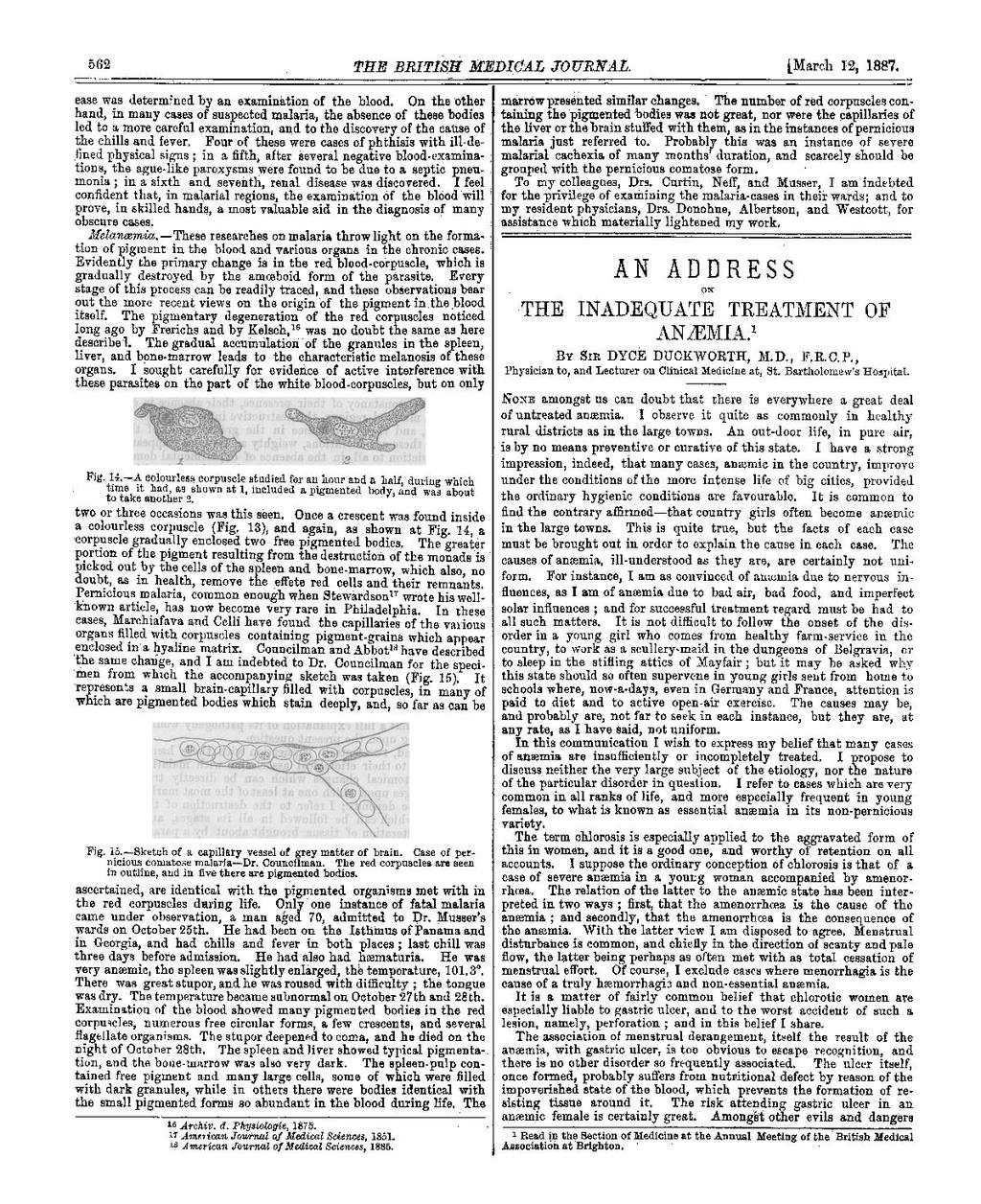
two or three occasions was this seen. Once a crescent was found inside a colourless corpuscle (Fig.13), and again, as shown at Fig.14, a corpuscle gradually enclosed two free pigmented bodies. The greater portion of the pigment resulting from the destruction of the monads is picked out by the cells of the spleen and bone-marrow, which also, no doubt, as in health, remove the effete red cells and their remnants. Pernicious malaria, common enough when Stewardson [2] wrote his well-known article, has now become very rare in Philadelphia. In these cases, Marchiafava and Celli have found the capillaries of the various organs filled with corpuscles containing pigment-grains which appear enclosed in a hyaline matrix. Councilman and Abbot[3] have described the same change, and I am indebted to Dr. Councilman for the specimen from which the accompanying sketch was taken (Fig.15). It represents a small brain-capillary filled with corpuscles, in many of which are pigmented bodies which stain deeply, and, so far as can be
ascertained, are identical with the pigmented organisms met within the red corpuscles during life. Only one instance of fatal malaria came under observation, a man aged 70, admitted to Dr. Musser's wards on October 25th. He had been on the Isthmus of Panama and in Georgia, and had chills and fever in both places; last chill was three days before admission. He had also had hæmaturia. He was very anemic, the spleen was slightly enlarged, the temperature, 101.3°. There was great stupor, and he was roused with difficulty; the tongue was dry. The temperature became subnormal on October 27th and 28th. Examination of the blood showed many pigmented bodies in the red corpuscles, numerous free circular forms, a few crescents, and several flagellate organisms. The stupor deepened to coma, and he died on the night of October 28th. the spleen and liver showed typical pigmentation, and the bone-marrow was also very dark. The spleen-pulp contained free pigment and many large cells, some of which were filled with dark granules, while in others there were bodies identical with the small pigmented forms so abundant in the blood during life. The marrow presented similar changes. The number of red corpuscles containing the pigmented bodies was not great, nor were the capillaries of the liver or the brain stuffed with them, as in the instances of pernicious malaria just referred to. Probably this was an instance of severe malarial cachexia of many months' duration, and scarcely should be grouped with the pernicious comatose form.
To my colleagues, Drs. Curtin, Neff, and Musser, I am indebted for the privilege of examining the malaria-cases in their wards; and to my resident physicians, Drs. Donohue, Albertson, and Westcott, for assistance which materially lightened my work.