and fuses with the supra-occipital. The opisthotic lies between the epiotic and the lateral occipital with which it ultimately fuses; in some birds, e.g. in Larus, it extends far enough to help to bound the foramen magnum. The basisphenoids are ventrally overlaid, and later on fused with, a pair of membrane bones, the basi-temporals, homologous in part with the parasphenoid of lower vertebrates. They contribute to the formation of the auditory meatus, and of the right and left carotid canals which accompany the eustachian tubes. In many birds the basisphenoids send out a pair of basipterygoid processes by which they articulate with the pterygoids. Dorso-laterally the basisphenoid is joined by the alisphenoid, which forms most of the posterior wall of the orbit. The orbito-sphenoids diverge only posteriorly, otherwise they are practically unpaired and form the median interorbital septum, which is very large in correlation with the extraordinary size of the eyeballs.
 |
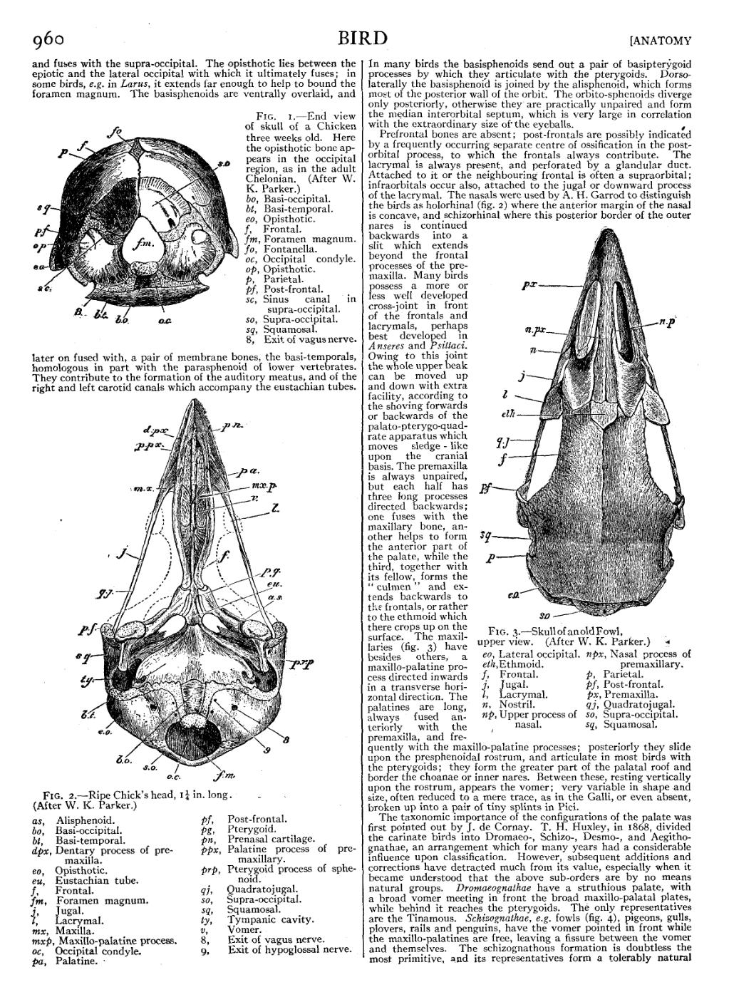
Fig. 1.—End view of skull of a Chicken three weeks old. Here the opisthotic bone appears in the occipital region, as in the adult Chelonian. (After W. K. Parker.) bo, Basi-occipital. bt, Basi-temporal. eo, Opisthotic. f, Frontal. fm, Foramen magnum. fo, Fontanella. oc, Occipital condyle. op, Opisthotic. p, Parietal. pf, Post-frontal. sc, Sinus canal in supra-occipital. so, Supra-occipital. sq, Squamosal. 8, Exit of vagus nerve. |

|
| Fig. 2.—Ripe Chick’s head, 114 in. long. (After W. K. Parker.) |
| bo, Basi-occipital. | pa, Palatine. |
| bt, Basi-temporal. | pf, Post-frontal. |
| dpx, Dentary process of premaxilla. | pg, Pterygoid. |
| eo, Opisthotic. | pn, Prenasal cartilage. |
| eu, Eustachian tube. | ppx, Palatine process of pre-maxillary. |
| f, Frontal. | prp, Pterygoid process of sphenoid. |
| fm, Foramen magnum. | qj, Quadratojugal. |
| j, Jugal. | so, Supra-occipital. |
| l, Lacrymal. | sq, Squamosal. |
| mx, Maxilla. | ty, Tympanic cavity. |
| mxp, Maxillo-palatine process. | v, Vomer. |
| oc, Occipital condyle. | 8, Exit of vagus nerve. |
| 9, Exit of hypoglossal nerve. |

| |
| Fig. 3.—Skull of an old Fowl, upper view. (After W. K. Parker.) | |
| eo, Lateral occipital. | npx, Nasal process of premaxillary. |
| eth, Ethmoid. | p, Parietal. |
| f, Frontal. | pf, Post-frontal. |
| j, Jugal. | px, Premaxilla. |
| l, Lacrymal. | qj, Quadratojugal. |
| n, Nostril. | so, Supra-occipital. |
| np, Upper process of nasal. | sq, Squamosal. |
Prefrontal bones are absent; post-frontals are possibly indicated by a frequently occurring separate centre of ossification in the post-orbital process, to which the frontals always contribute. The lacrymal is always present, and perforated by a glandular duct. Attached to it or the neighbouring frontal is often a supraorbital; infraorbitals occur also, attached to the jugal or downward process of the lacrymal. The nasals were used by A. H. Garrod to distinguish the birds as holorhinal (fig. 2) where the anterior margin of the nasal is concave, and schizorhinal where this posterior border of the outer nares is continued backwards into a slit which extends beyond the frontal processes of the premaxilla. Many birds possess a more or less well developed cross-joint in front of the frontals and lacrymals, perhaps best developed in Anseres and Psittaci. Owing to this joint the whole upper beak can be moved up and down with extra facility, according to the shoving forwards or backwards of the palato-pterygo-quadrate apparatus which moves sledge-like upon the cranial basis. The premaxilla is always unpaired, but each half has three long processes directed backwards; one fuses with the maxillary bone, another helps to form the anterior part of the palate, while the third, together with its fellow, forms the “culmen” and extends backwards to the frontals, or rather to the ethmoid which there crops up on the surface. The maxillaries (fig. 3) have besides others, a maxillo-palatine process directed inwards in a transverse horizontal direction. The palatines are long, always fused anteriorly with the premaxilla, and frequently with the maxillo-palatine processes; posteriorly they slide upon the presphenoidal rostrum, and articulate in most birds with the pterygoids; they form the greater part of the palatal roof and border the choanae or inner nares. Between these, resting vertically upon the rostrum, appears the vomer; very variable in shape and size, often reduced to a mere trace, as in the Galli, or even absent, broken up into a pair of tiny splints in Pici.
The taxonomic importance of the configurations of the palate was first pointed out by J. de Cornay. T. H. Huxley, in 1868, divided the carinate birds into Dromaeo-, Schizo-, Desmo-, and Aegithognathae, an arrangement which for many years had a considerable influence upon classification. However, subsequent additions and corrections have detracted much from its value, especially when it became understood that the above sub-orders are by no means natural groups. Dromaeognathae have a struthious palate, with a broad vomer meeting in front the broad maxillo-palatal plates, while behind it reaches the pterygoids. The only representatives are the Tinamous. Schizognathae, e.g. fowls (fig. 4), pigeons, gulls, plovers, rails and penguins, have the vomer pointed in front while the maxillo-palatines are free, leaving a fissure between the vomer and themselves. The schizognathous formation is doubtless the most primitive, and its representatives form a tolerably natural