the bone has three surfaces for the attachment of muscles, and a sharp outer border for the interosseous membrane. The lower end, much smaller than the upper, has a pointed styloid process and a smooth articular surface, the outer portion of which is for the lower end of the radius, the lower part for moving on a cartilage of the wrist joint called the triangular fibro-cartilage.
The hand consists of the carpus or wrist, of the metacarpus or palm, and of the free digits, the thumb and four fingers. Anatomists H . describe it with the palm turned to the front, and with its
axis in line with the axis of the forearm. The carpal or wrist bones (fig. 14) are eight in number and small in size : they are arranged in two rows, a proximal, — i.e. a row Carous next the forearm, — consisting of the scaphoid, semilunar, ' cuneiform and pisiform ; and a distal, — i.e. a row next the bones of the palm, — consisting of a trapezium, trapezoid, os magnum and unciform; the bones in each row being named in the order they are met with, from the radial or outer to the ulnar or inner side of the wrist. It is unnecessary to give a separate de- scription of each bone. Except the pisiform or pea-shaped bone, which articulates with the front of the cuneiform, each carpal bone is short and irregularly cuboidal in shape; its anterior (or palmar) surface and its posterior (or dorsal) being rough, for the attachment of ligaments; its superior and inferior surfaces being invariably smooth, for articulation with adjacent bones; whilst the inner and outer surfaces are also smooth, for articulation, except the outer surfaces of the scaphoid and trapezium (the two external bones of the carpus), and the inner surfaces of the cuneiform and unciform (the two internal bones). Occasionally extra bones are found, but they are apparently the remnants of cartilaginous elements found in the hand of the early embryo (see G. Thilenius, Morph. Arbeiten, v., 1896).
The metacarpal bones, or bones of the palm of the hand, are five in number (fig. 14). They are miniature long bones, and each possesses a shaft and two extremities. The metacarpal of the thumb is the shortest, and diverges outward from the rest; its carpal extremity is saddle-shaped, for articulation with the trapezium ; its shaft is somewhat compressed, and its phalangeal end is smooth and rounded, for the first phalanx of the thumb. The four other metacarpal bones belong to the four fingers ; they are almost parallel to each other, and diminish in size from the second to the fifth. Their carpal ends articulate with the trapezoid, os magnum and unciform: their shafts are three-sided: their phalangeal ends articulate with the proximal phalanges of the fingers.
The number of digits in the hand is five. They are distinguished by the names of pollex or thumb, index, medius, annularis and _. „ minimus. Their skeleton consists of fourteen bones,
uigns. named phalanges, of which the thumb has two, and each
of the four fingers three. The phalanx next the metacarpal bone is the proximal, that which carries the nail, the terminal or ungual phalanx, whilst the intermediate bone is the middle phalanx. Each is a miniature long bone, with two articular extremities and an intermediate shaft, except the terminal phalanges, which have an articular surface only at their proximal ends, the distal end being rounded and rough, to afford a surface for the lodgment of the nail.
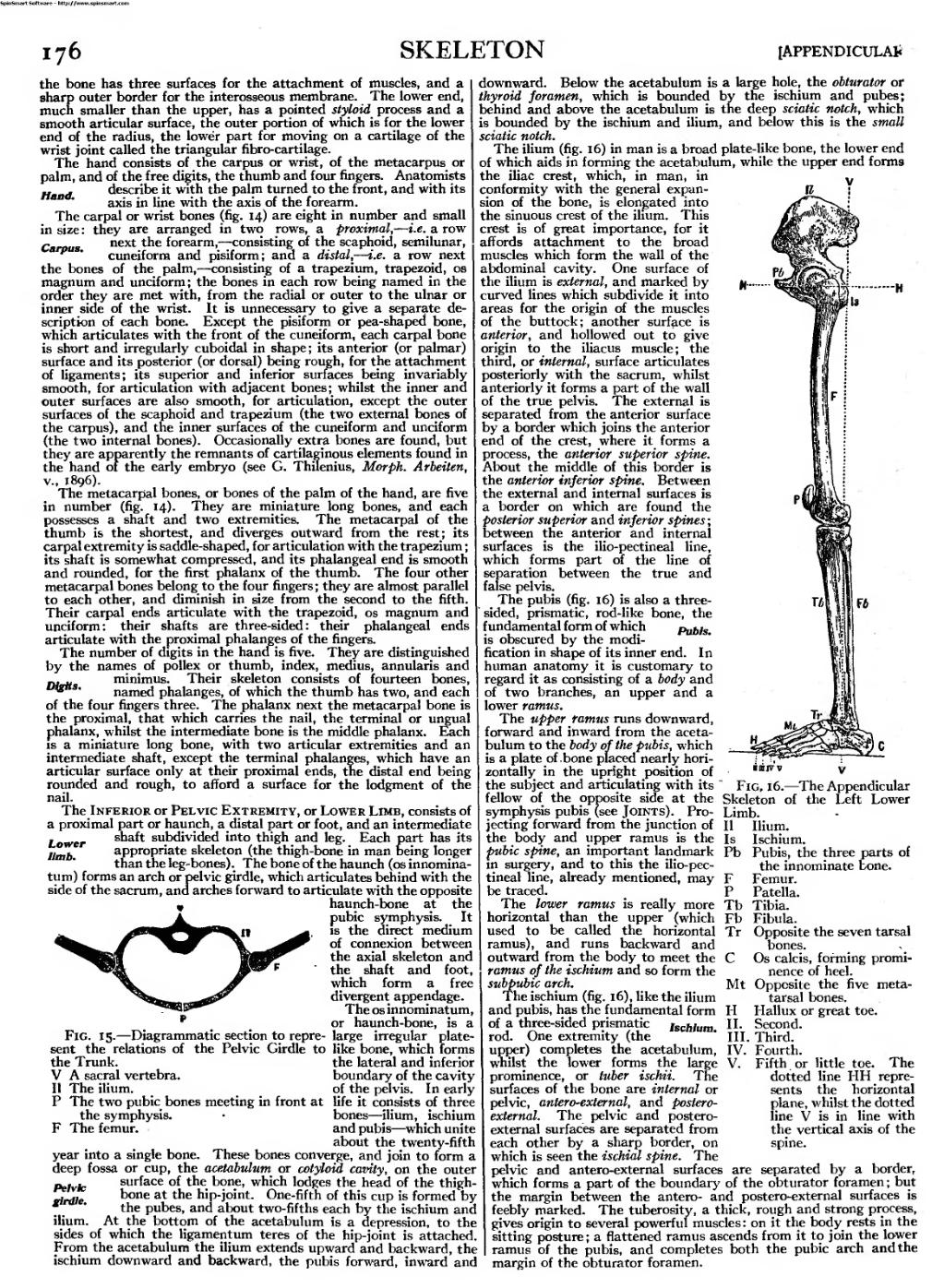
The Inferior or Pelvic Extremity, or Lower Limb, consists of a proximal part or haunch, a distal part or foot, and an intermediate shaft subdivided into thigh and leg. Each part has its appropriate skeleton (the thigh-bone in man being longer than the leg-bones). The bone of the haunch (os innomina- tum) forms an arch or pelvic girdle, which articulates behind with the side of the sacrum, and arches forward to articulate with the opposite
haunch-bone at the pubic symphysis. It is the direct medium of connexion between the axial skeleton and the shaft and foot, which form a free divergent appendage.
The os innominatum, or haunch-bone, is a Fig. 15. — Diagrammatic section to repre- large irregular plate- sent the relations of the Pelvic Girdle to like bone, which forms
Lower limb.
downward. Below the acetabulum is a large hole, the obturator or
thyroid foramen, which is bounded by the ischium and pubes;
behind and above the acetabulum is the deep sciatic notch, which
is bounded by the ischium and ilium, and below this is the small
sciatic notch.
The ilium (fig. 16) in man is a broad plate-like bone, the lower end of which aids in forming the acetabulum, while the upper end forms the iliac crest, which, in man, in conformity with the general expan- sion of the bone, is elongated into the sinuous crest of the ilium. This crest is of great importance, for it affords attachment to the broad muscles which form the wall of the abdominal cavity. One surface of the ilium is external, and marked by curved lines which subdivide it into areas for the origin of the muscles of the buttock; another surface is anterior, and hollowed out to give origin to the iliacus muscle; the third, or internal, surface articulates
posteriorly with the sacrum, whilst anteriorly it forms a part of the wall of the true pelvis. The external is
separated from the anterior surface
by a border which joins the anterior
end of the crest, where it forms a
process, the anterior superior spine.
About the middle of this border is
the anterior inferior spine. Between
the external and internal surfaces is
a border on which are found the
posterior superior and inferior spines;
between the anterior and internal
surfaces is the ilio-pectineal line,
which forms part of the line of
separation between the true and
false pelvis.
The pubis (fig. 16) is also a three- sided, prismatic, rod-like bone, the
fundamental form of which Pubis.
is obscured by the modi- fication in shape of its inner end. In
human anatomy it is customary to
regard it as consisting of a body and
of two branches, an upper and a
lower ramus.
The upper ramus runs downward,
forward and inward from the aceta- bulum to the body of the pubis, which
is a plate of bone placed nearly hori- zontally in the upright position of ■«ff» v
the subject and articulating with its " Fig, 16. — The Appendicular
fellow of the opposite side at the Skeleton of the Left Lower
symphysis pubis (see Joints). Pro- Limb.
jecting forward from the junction of II Ilium.
the body and upper ramus is the Is Ischium.
the Trunk.
V A sacral vertebra.
II The ilium.
P The two pubic bones meeting in front at
the symphysis. F The femur.
the lateral and inferior
boundary of the cavity
of the pelvis. In early
life it consists of three
bones — ilium, ischium
and pubis — which unite
about the twenty-fifth
year into a single bone. These bones converge, and join to form a
deep fossa or cup, the acetabulum or cotyloid cavity, on the outer
Pelvh surface of the bone, which lodges the head of the thigh-
rlrdle ^one at the hip-joint. One-fifth of this cup is formed by
â„¢ ' the pubes, and about two-fifths each by the ischium and
ilium. At the bottom of the acetabulum is a depression, to the sides of which the ligamentum teres of the hip-joint is attached. From the acetabulum the ilium extends upward and backward, the ischium downward and backward, the pubis forward, inward and
fubic spine, an important landmark Pb Pubis, the three parts of in surgery, and to this the ilio-pec-
tineal line, already mentioned, may be traced.
The lower ramus is really more horizontal than the upper (which used to be called the horizontal ramus), and runs backward and outward from the body to meet the ramus of the ischium and so form the subpubic arch.
The ischium (fig. 16), like the ilium and pubis, has the fundamental form of a three-sided prismatic ischium. rod. One extremity (the upper) completes the acetabulum, whilst the lower forms the large V. prominence, or tuber ischii. The surfaces of the bone are internal or pelvic, antero-external, and postero- external. The pelvic and postero
the innominate bone.
Femur.
Patella.
Tibia.
Fibula.
Opposite the seven tarsal bones.
Os calcis, forming promi- nence of heel.
Opposite the five meta- tarsal bones.
Hallux or great toe.
Second.
Third. IV. Fourth.
Fifth or little toe.
dotted line HH repre- sents the horizontal plane, whilst the dotted line V is in line with the vertical axis of the spine.
Mt
H
II.
III.
The
external surfaces are separated from each other by a sharp border, on which is seen the ischial spine. The pelvic and antero-external surfaces are separated by a border, which forms a part of the boundary of the obturator foramen ; but the margin between the antero- and postero-external surfaces is feebty marked. The tuberosity, a thick, rough and strong process, gives origin to several powerful muscles : on it the body rests in the sitting posture ; a flattened ramus ascends from it to join the lower ramus of the pubis, and completes both the pubic arch and the margin of the obturator foramen.