Popular Science Monthly/Volume 79/December 1911/Protozoan Germ Plasm
| PROTOZOAN GERM PLASM |
By Professor GARY N. CALKINS
COLUMBIA UNIVERSITY
IN his classical essays on the nature of the germ plasm, Weismann, more than twenty years ago, drew a distinction between that protoplasm destined for the perpetuation of the race and that needed by the organism for its ordinary functions of moving, eating, digesting, etc. The former, which he designated germ plasm, in Metazoa is early differentiated from the latter, and in some forms may be distinguished as the rudiments of a germinal epithelium even before the end of segmentation. The latter develops into the vegetative organs of the adult and serves to nourish and support the former. The distinction, therefore, especially in the higher Metazoa, indicates a real difference in potential, and the vegetative cells have no primary reproductive functions. In lower Metazoa the distinction is not so clear, many of the vegetative cells turning to germ cells either in groups or singly.
Protozoa, or animals consisting of one cell only, were set apart by Weismann as differing from Metazoa in not showing this somatic germinal differentiation, and he regarded them all as potential germ cells. Furthermore, since germ cells have the possibility, at least, of continued life, while somatic cells die, he assumed that Protozoa are potentially immortal, while natural death is the penalty Metazoa must pay for the privilege of differentiation.
Weismann's hypothesis is certainly seductive, and, viewed superficially, would seem to indicate a fundamental difference between the unicellular and the multicellular animals. Protozoa, however, are more than mere single cells, comparable with the isolated tissue cells of higher animals. They must be regarded as organisms, complete in themselves and comparable, therefore, with the whole animal of higher type and not with any one of its cells. Like the entire Metazoon, it excretes the products of destructive metabolism, it secretes many different types of by-products; it moves, obtains food, swallows, digests and assimilates it through the action of digestive fluids; in short, it performs all of the functions which distinguish animals from plants. Finally, like higher types again it reproduces its kind by processes relatively as simple as the functions of digestion or nervous response are simple when compared with these functions in Metazoa. In such complete organisms, therefore, it is a logical inference to consider the protoplasm of a protozoon as made up of widely different elements equivalent in function to the aggregate of cells making up the metazoon, and with some parts at least having the power to contract and move, some to digest food, some to secrete, others to excrete and still others, finally, to reproduce. Considered in this sense the cell theory as applied  Fig. 1.Original. to the Protozoa is obviously inadequate.
The especial portions of the protoplasm that have to do with these several different functions of the protozoon can be identified in many cases as structurally different from the remainder, especially those parts which have to do with movement and with the perpetuation of the race, i. e., the germ plasm. In the majority of Protozoa this portion of the protozoon is clearly differentiated, one time or another, from the remainder of the cell, and this justifies us in taking issue with Weismann, and with the majority of those who write casually about the Protozoa, as having no somatic cell elements and therefore no possibility of natural death. If it can be shown that there is a specific germ plasm in these unicellular animals, then the matter of immortality differs in no essential way from the same problem in Metazoa or Metaphyta where the germ cells have a possibility of endless existence. The evidence points to a common and universal law that continued life is an attribute of an especially endowed protoplasm, termed germ plasm, which forms the material basis of the reproductive cells.
The history of single Protozoa, if taken superficially, seems to point to the fact that the protoplasm of these cells does not die a natural death, but continues to live in successive generations of similar individuals. The individual cell appears to be entirely self-sufficient; it captures food, digests it, grows to maximum size and then divides (Fig. 1). The original individual cell no longer exists, although it has not died; its protoplasm is now distributed between two daughter cells. In the same way these cells grow old in turn, divide into two each, and so on apparently in endless succession of cell generations. Obviously if this could keep on indefinitely there would be a basis for the view that Protozoa are immortal. They do not keep this up, however, but there comes a time when the nature of the protoplasmic make-up changes, and processes similar to fertilization in Metazoa supervene. In the great majority of parasitic protozoa and in most free-living  Fig. 2.Original. forms that have been studied in culture, there comes a period when certain cells of the race, or specialized parts of the protoplasm of all of the cells of the gig race, undergo marked changes. different from any vegetative phase, and reorganization of the old individual or formation of new ones is the outcome. This result is brought about by conjugation or the union of two cells in more or less complete coalescence, during which an interchange and mixture of germ plasms is accomplished (Fig. 2).
cells which now undergo, separately, the later stages of development. These stages consist in the division of the fertilization nucleus and new formation from the division products, of the new macronucleus and the new micronucleus (Fig. 4).
After such a process of fertilization it would seem that the individuals are pretty much as they were before except for the complete reorganization of the nuclear apparatus, and there is a certain justification for the Weismann conception. But the phenomena in Paramecium and allied forms, like their cell differentiations, are highly specialized and are unlike the fertilization processes in the majority of Protozoa.
The enormous group of Sarcodina, including more than four thousand species of Radiolaria and some thousand or more species of Foraminifera, Heliozoa and Rhizopods, presents a fairly uniform picture of the germ plasm and the processes of fertilization. For purposes of illustration and comparison I will describe two types selected from this great group of forms—one a marine foraminiferon, Polystomella, crispa, the other a common fresh-water rhizopod Arcella vulgaris.
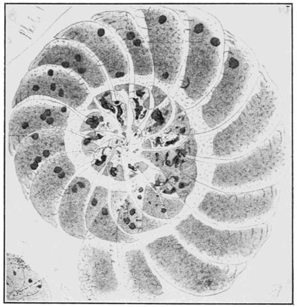
So far as known, each species of Foraminifera exists in two forms known as the microsphæric and the megalosphæric forms, so called because of the small and large size of the central or initial chamber of the shell (Figs. 5 and 6). These two forms correspond with the asexual and the sexual generations of metagenetic hydrozoa, the microsphæric type corresponding with the hydroid, the macrosphæric type with the medusa generation. Like these cœlenterates, the microsphæric type reproduces asexually while the macrosphæric type reproduces sexually. Like them also, the asexual generation gives rise to the sexual and the latter, again, to the asexual, hence there is a typical alternation of generations. Like the cœlenterates, again, the sexual generation acts as a nurse for the important germ plasm. Let us see how this works out in the case of Polystomella crispa.
The young individual of Polystomella secretes a shell of calcium carbonate and grows by feeding on various minute animals and plants. Its nucleus divides by mitosis and the protoplasmic mass increases in size but does not divide with the nucleus. A new shell chamber is formed partly enclosing the first one. Further division of the nuclei, increase of the plasmic mass and new chamber formation continues with constant feeding until a typical Polystomella shell is formed, containing a relatively great protoplasmic mass and hundreds of nuclei. When mature, all of the nuclei save one or two break down into thousands of minute particles of chromatin which are distributed throughout the protoplasm in the form of fine granules. Authorities differ somewhat in regard to the next changes of the nuclei. If we accept Schaudinn's
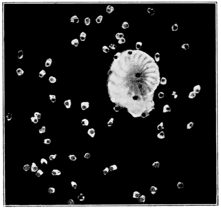
account, verified by Winter, the protoplasm of the microsphæric individual breaks down into small fragments, each fragment enclosing a number of these granules of chromatin, which coalesce later to form one single nucleus. If we accept Lister's account the coalescence occurs before the breaking down of the protoplasmic mass. All observers agree, however, that hundreds of minute nucleated "embryos" arise
Fig. 7. Polystomella crispa. Liberation of pseudo-podiospores from the megalosphæric individual.
by fragmentation of the parent protoplasm (Fig. 7). The breaking down of the original nuclei is an important step, for by this process the important germ plasm is formed, which in the finely granular state described was named by Hertwig the chromidia.
The small "embryos" leave the parent organism in swarms (Fig. 8), and the calcareous shell is finally deserted. Each "embryo" then grows into a megalosphæric or large-chambered individual in which the primary nucleus remains single for a considerable period. New chambers are formed about the primary central chamber and a new adult individual results from the continued growth. The history of the nucleus, however, is quite different from that of the microsphæric individual. The protoplasm contains a large primary nucleus which ultimately degenerates and disappears. During life of the individual, however, this nucleus gives rise by chromatin "secretion" or by fragmentation, to an immense number of minute nuclei which are distributed throughout the protoplasmic mass. Each nucleus becomes surrounded by a thickened zone of protoplasm and then each divides twice, thus increasing the number of the zones. All of the protoplasm is utilized in this zone formation, with the exception of a small portion surrounding the original "primary" nucleus, these parts degenerating and disappearing. Preparations of the decalcified individual at this period show hundreds of minute nucleated masses completely filling the shell space (Fig. 6). In life, these emerge in swarms, each in the form of a tiny bi-flagellated swarmer. The swarmers are gametes which
conjugate with similar gametes from another individual (Fig. 8, A). The flagella are thrown off after union, the nuclei unite, and each united pair, as a fertilized cell, or zygote, develops into a new microsphæric individual.
In Polystomella, therefore, fertilization is accomplished, not by union of the parent individuals as in Paramecium, but by coalescence and fusion of minute gametes which contain portions of the specific germ substance in the form of chromidia.
Arcella vulgaris, a common fresh-water shelled rhizopod, has a much more simple life-history. The nucleus of the young form soon divides, so that most Arcella specimens contain  Fig. 9.Original. two primary nuclei (Fig. 9). From the very outset, furthermore, each nucleus secretes a chromatin substance which collects in a zone about the periphery of the cell. This substance is not granular like the chromidia, but has a similar origin from the nucleus, and has the same germ-plasmic fate as chromidia, so that Hertwig was justified in calling it a "chromidial net." When the organism is mature, minute nuclei condense out of the substance of this network, hundreds of them being formed (Fig. 10). As in Polystomella, each nucleus becomes surrounded by a zone of protoplasm, and, finally, a large number of small swarmers emerge from the shell mouth, leaving behind in the shell the two primary nuclei and a portion of the protoplasm as a degenerating residue. The swarmers are dimorphic,
some are macrogametes, some microgametes (Fig. 11). These fuse two by two, a macrogamete with a microgamete, and the resulting zygote develops into the normal form.
In Arcella, therefore, as in Polystomella, there is a definite germinal protoplasm composed essentially of chromatin which gives rise to new cycles of organisms, while a portion of the original organism, including the primary nucleus and a quantity of protoplasm, degenerates and dies. We can speak of germinal and somatic protoplasm in these cases, and  Fig. 11.After Elpetiewsky with equal right in all of the thousands of allied species of protozoa, as well as in the case of any higher animal.
A still more interesting case of specific germ-plasm formation is given by a type of Gregarine belonging to a group of parasitic Sporozoa. In all of the Gregarines there is a segregation of the germ plasm and a residual somatic protoplasm which degenerates and dies a somatic death. One of the most striking illustrations of this type is the case of Ophryocystis mesnili, a parasite of beetles. Unlike the cases cited above, most of the gregarines do not reproduce asexually. Ophryocystis, however, is one of the exceptions to the rule, the individuals reproducing by simple division until the protoplasm becomes mature, when, as in Paramecium, two cells come together in conjugation. The single nucleus of each cell divides and one of the products becomes a "somatic" nucleus, to use Leger's term, while the other daughter nucleus in each cell divides again (Fig. 12, A-G). One of these corresponds to a polar body in the metazoon egg, the other is the gamete, or germ, nucleus. In each cell this nucleus collects about itself, possibly through the secretion of nuclear material which transforms the surrounding stuff, a denser zone of protoplasm, which, with the nucleus, forms a gamete within the body of the parent cell. The two gametes thus formed fuse while in the space which their formation has left in the parent somatic, or nurse cells. The latter ultimately wither up and die. After union of the two gametes, the sporoblast gives rise to eight germs or sporozoites, each capable of developing into an ordinary vegetative form when under the proper conditions of environment (Fig. 12, H-N).
Here in Gregarines, therefore, as in rhizopods, we see a clearly defined difference between germ plasm and somatic plasm, the latter dying, as in the Metazoa, the former capable of endless development. Unlike the rhizopods, however, the germinal chromatin is retained in the primary nucleus until full maturity of the cell and does not appear in the cytoplasm in the form of chromidia.
Turning after this excursion into other fields of Protozoa, to the case of Paramecium, it will be seen that the conditions are entirely different from those which accompany conjugation in the majority of
Protozoa. In the latter fertilization is accomplished by the formation and fusion of gametes or minute simulacra of the adult cells, some being differentiated into microgametes and macrogametes, or male and female. In Paramecium there are no gametes of this kind, but portions of the adult individuals fuse as in Ophryocystis mesnili. Unlike this Gregarine, the fusion of the adult cells is only temporary and the two parties to the conjugation do not die. During the temporary fusion there is a mutual interchange of micronuclei and a mutual fertilization, while the only portion to disintegrate and die is the macronucleus of each conjugant, and this is replaced by a specially differentiated fragment of the new micronucleus.
In ciliated protozoa such as Paramecium and its near relations the germ plasm is concentrated in the micronucleus, while the somatic plasm is represented by the macronucleus. As we have seen, the micronucleus enlarges and divides during conjugation, first into two, then these into four, all but one of the four degenerating. The fourth divides for the third time, but this time unequally (Fig. 3) into a smaller migratory and a larger stationary form. The smaller micronucleus migrates into the other cell of the pair and there unites with the stationary larger nucleus. The macronucleus then degenerates and is absorbed in each of the conjugating cells.
The aberrant conditions in these Infusoria can be interpreted if we regard the three divisions of the micronucleus as the reminiscence of a
process of gamete formation which obtained in remote ancestral forms. A parallel, but less extreme, case is seen in the Gregarine Ophryocystis, where one gamete only is formed by the conjugating cells. This is an isolated case among these Sporozoa, for in the typical forms a great number of gametes are developed, as shown in the accompanying photograph of Monocystis (Fig. 13). The one pair of gametes in Ophryocystis, plus the nuclear derivatives of the pro-conjugants, represent the swarm of gametes found in other Gregarines. Here, also, the internal or "nursed"; condition of the gametes is a new development or adaptation.
Similarly in Paramecium the three divisions of the micronucleus may be interpreted as representing an ancestral brood of conjugating gametes, only two of which are now functional, the one representing a macrogamete or female form, the other a microgamete or male cell. Unlike Ophryocystis, the Paramecium individual does not become a nurse for the conjugating gametes, but remains, as before, the mechanism for the performance of the various physiological activities and the vehicle of the somatic and germ plasms.
The widely accepted view, therefore, as first formulated by Weismann and repeatedly stated in general works on biology, that Protozoa differ from Metazoa in having no equivalent of the somatic cells and therefore no somatic, or natural, death, must be abandoned. In the vast majority of Protozoa there is a clearly defined equivalent of somatic cells and an equivalent of natural death. The conditions in Paramecium and its allies are different from those of other protozoa, the old individual does not die at conjugation but is completely reorganized and built up of parts derived from the product of fertilization exactly as in Metazoa. The protozoon is not a potential germ cell, but like the metazoon is the carrier of the racial germ plasm which, in the great majority of protozoa is differentiated from the somatic plasm. As the germ cells of the metazoon become segregated into a germinal epithelium, becoming functional at maturity, so the germ plasm of the protozoon becomes segregated as chromidia or granules of a specific kind of chromatin, in the cell and is likewise functional at maturity.


