Popular Science Monthly/Volume 84/May 1914/The Laboratory of Comparative Pathology of the Zoological Society of Philadelphia
| THE LABORATORY OF COMPARATIVE PATHOLOGY OF THE ZOOLOGICAL SOCIETY OF PHILADELPHIA |
By R. W. SHUFELDT, M.D.
WASHINGTON, D. C.
IN various publications recently I have pointed out the fact as to how little is being done in the way of describing the anatomy of the existing Vertebrata of our fauna. One animal after another is now being exterminated with a rapidity never before equalled in the history of man, neither has there ever been a time in that history when so little was done to preserve detailed accounts, properly illustrated, of the comparative morphology of the species so doomed.
This state of things is not entirely confined to our own country by any means, for the same neglect is but too apparent elsewhere. Faunas are being exterminated and material recklessly wasted at zoological gardens, laboratories and other places to an extent that is most deplorable. Comparative anatomists of the next century will be fully justified in saying what they please of such criminal neglect as this, when they come to realize the extent to which those of the present one ignored their opportunities in this field of scientific research, and allowed so many animals to die out without leaving the shadow of a record describing their structure.
We are doing much better with respect to the study of the causes of death in those ferine forms which die in captivity, for the activity along such lines is very marked and more or less universal. Not only are the diseases of the vertebrates below man being studied in numerous and fully equipped institutions in this country and abroad, but, through various scientific methods, comparative pathology, including that of man and the domesticated animals, is being investigated, studied and utilized in a manner far more extensive than has ever been the case in the history of our race. Such investigations include the parasites of the Vertebrata, a field of research which has received so much attention during recent years at the hands of Dr. F. E. Beddard, prosector of the Zoological Society of London, in the Old World, and Dr. Charles Wardell Stiles, of the Bureau of Animal Industry of this country.
Recently I have been in communication with Dr. Herbert Fox on this subject, and he has kindly placed at my disposal a set of photographs illustrating the building and the work rooms of the Laboratory of Comparative Pathology of the Zoological Society of Philadelphia, of which institution he is now the pathologist in charge. Dr. Fox has also

sent me some excellent photographs of pathological specimens prepared at his laboratory and obtained from animals which have died at the Philadelphia Zoological Garden. These are very instructive, indeed; but, much as I regret the fact, they can not well he used in the present connection.
It was in 1901 that pathological work upon the animals that died at the Zoo was inaugurated, and this at the instance of Dr. Charles B. Penrose, of Philadelphia, who had as advisers in the matter the late Dr. Leonard Pearson, of the Pennsylvania Veterinary Department, and Dr. M. P. Ravenel, of the State Stock Sanitary Board.
The first pathologist to the garden was Dr. C. Y. White, assistant director of the William Pepper Laboratory of Clinical Medicine, in which laboratory the post mortems were done.
We learn from the Thirty-first Annual Report of the Board of Directors of the Zoological Society (1903) that no fewer that seventy-six different species of mammals had there been examined with the view of ascertaining the cause of their death, and the result of the autopsies recorded (pp. 20-25). A large number of these mammals were various species of monkeys, apes and their allies (Primates), and the great majority of these succumbed to general tuberculosis.
Besides this much-dreaded malady, these animals suffered from twenty-five other diseases of which they were the victims; this does not
include the parasites observed, nor one bird—a cow bird—in which case molluscum contagiosum proved fatal. These examinations were made during the period from November, 1901, to March 1, 1903. Numerous other animals also died during this time of which no record was kept and no examination made; these were principally birds and indigenous species of mammals, reptiles and other forms. They died chiefly from the injuries and disturbances incident to their capture and captivity, most often shortly after reception, and, as a general thing, no special use was ever made of them.
Dr. Penrose reported that
In ten of the animals examined no cause of death was discovered, though careful investigation of all the organs was made, as well as bacteriological examination of the blood. Change in food, water, temperature and general environment, may cause the death of wild animals in captivity, without producing gross or apparent lesions of any of the structures of the body. It is probable that in some cases the animal dies of a toxæmia due to improper food, though we have been unable to determine the existence of this condition at autopsy. The post mortem changes have usually rendered the bacteriological examination very unsatisfactory.
These reports of Drs. Penrose and White, continuing until 1906, are of great interest and importance, but altogether too extensive even for summarization here.
In 1905 a building was selected upon the grounds of the garden and, at some expense, remodeled for a pathological laboratory, Dr.
Fox taking charge of this the following year (Fig. 1). As the operations of this department became more extensive, it was realized that the labor of no one man could properly compass it; therefore, in 1910, Dr. Fred D. Weidman, who at the time was assistant in the department of pathology at the University of Pennsylvania, received the appointment of assistant pathologist, and the following year a second floor was put upon the building to accommodate the museum specimens and for additional work-room.
From about this time on, the principal part of the report of the society was given over to that of the pathological laboratory, a most gratifying indication of the value of the work performed there. As evidence of this we find that the thirty-ninth annual report of the board of directors of the society (1911) prints forty-seven small octavo pages, and of these Dr. Fox's report of the laboratory occupies from pages 15 to 40 inclusive.
Far-reaching in its importance, this report contains most valuable data, which can not fail to be of use, not only to keepers of gardens and menageries, but to the human pathologist and the breeder of domestic animals. The report goes to show that no fewer than 325 animals were examined, of which number 93 were mammals. There is an excellent report on "Tuberculin Reaction in Monkeys" containing much of practical value, while what Dr. Weidman gives on "Parasites" is not only quite extensive, but presented in great detail. A valuable special report
on "Bird Diphtheria" follows, with others on "Recurrent Ophthalmia," and one made by Drs. E. A. Schumann and Fox on "Leucocyte Counts." All these are in evidence of the marked activity of this very efficient laboratory, and the character of the contributions we may expect from it in the future. Autopsies are performed on all the animals which die in the gardens, with the exception of the small reptiles. Dr. Fox says:
The data concerning the animal during life is sent to the laboratory from the office on a special card form and the pathological findings are put upon this card. It is then used as a zoological index card and the diagnoses are cross indexed in a pathological system. Routine and special pathological and bacteriological methods are used as in the usual laboratory systems. The data obtained from these diagnoses is used in the hygiene of the garden and for scientific record.
Included in the laboratory 's work is the testing of animals suspected of having tuberculosis, a test made by subcutaneous injection of tuberculin. All monkeys received by the garden are observed for several days, and a record made of their daily 3 p. m. temperature. They are then tested, and if negative to this test are passed to the general monkey collection. If the test be doubtful, they are either held in quarantine or put on exhibition in isolated cages. If the test be positive they are killed. In order to avoid the carrying of tubercle bacilli on the hair they are washed in phenol solution upon arriving in and leaving the laboratory. Because of the frequent occurrence of proventricular worms in the parrots, the laboratory also examines the excrement of all new arrivals before they are put on exhibition.
This laboratory consists of an equipment of a two-floored building. We find on the first floor the general laboratory workroom (Fig. 2),
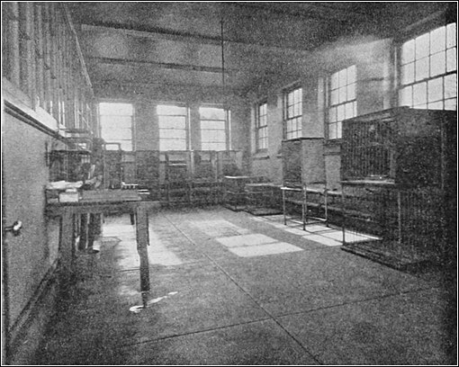
fitted up for special experiments and investigations. On the same floor there are two quarantine rooms, one of which is here shown in Fig. 3, wherein aie seen the cages in which are kept the animals under observation, or those presenting pathological conditions requiring their isolation. Beyond these we have the autopsy room, fitted out with all the modern appliances for performing post mortems.
In the case of three of these rooms, the walls are coated with hard paint, and center-drains are found in their concrete floors for the purpose of frequent flushing. One of the quarantine cages measures 27 24 20 inches and a larger one 29 36 30 inches, the top in any case being five feet from the floor. These comfortable quarters are of galvanized iron and fitted with a door which can not be opened by the animal.
Passing to the second floor of this building, we find it given over to a single room of considerable size, measuring thirty feet by sixty-nine. It is lighted overhead by a large, central skylight, while windows are only found in the north and east walls, the south and west ones being unpierced in any way to admit light. In this room is kept the collection of pathological specimens, some of which are from human subjects for the purposes of comparison. Many drawings, charts and photographs are upon the walls, while tables and desks are placed in convenient corners for the use of those doing clerical or laboratory desk work. For the purpose of destroying the animal remains after autopsies and dissections, there is in the building a direct incinerating plant—a most important adjunct to an institution of this kind.
The personnel of this laboratory is not large, being at present confined to the pathologist, assistant pathologist, technical assistant and a diener.
Should it come to pass at any time later on that the pathological work now being prosecuted in this very efficient department of the Philadelphia garden be supplemented by similar researches upon normal anatomy, for the purpose of which plenty of material is constantly there at hand, it will become necessary to increase the number on the present staff by adding to it a duly qualified prosector, zoological artist, photographer, and one or two additional assistants. It is very much to be hoped that this will be brought about in the near future.


