File:Steven H3N2 Flu ET.jpg
Steven_H3N2_Flu_ET.jpg (400 × 400 pixels, file size: 142 KB, MIME type: image/jpeg)
| This is a file from the Wikimedia Commons. Information from its description page there is shown below. Commons is a freely licensed media file repository. You can help. |
Summary
| Description |
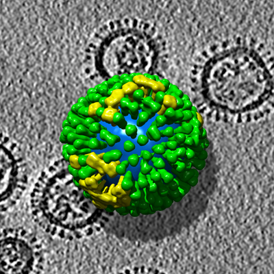
Afrikaans: Hierdie driedimensionele struktuur van H3N2-stam van Griepvirus-A is in 2006 met elektrontomografie verkry. Die virus is sowat 120 nanometer, dit is omtrent een tienduisendste van 'n millimeter, in deursnee.
Molekulêre Anatomie van Influensavirus Ontleed Wetenskaplikes by die Nasionale Instituut vir Artritis, Muskuloskeletale en Velsiektes (NIAMS), 'n afdeling van die Nasionale Institute vir Gesondheid in Bethesda, Maryland, en kollegas verbonde aan die Universiteit van Virginia in Charlottesville het daarin geslaag om, in ongekende detail, die virus uit te beeld wat griep veroorsaak. 'n Span navorsers onder leiding van NIAMS se Alasdair Steven, Ph.D., wat met 'n weergawe van die seisoenale H3N2-stam van griep A-virus werk, kon vyf verskillende soorte griepviruspartikels in dieselfde isolaat (monster) onderskei, om dan die molekule-verspreiding in elkeen te kon karteer. Hierdie deurbraak het die potensiaal om spesifieke kenmerke van hoogs virulente stamme te identifiseer, en om insig te verskaf in die wyse wat teenliggaampies die virus deaktiveer, en hoe virusse vatbare selle herken en hulle tydens infeksie binnedring. "Visualisering van influensaviruspartikels behoort 'n hupstoot te gee aan ons pogings om vir 'n moontlike pandemiese griepuitbraak voor te berei," sê NIAMS-direkteur Stephen I. Katz, M.D., Ph.D. “Hierdie werk sal ons in staat stel om ons vyand baie beter te ken.” Een van die probleme wat strukturele studies van die griepvirus belemmer het, is dat geen twee viruspartikels dieselfde is nie. In hierdie fundamentele opsig verskil dit van ander virusse. Die poliovirus, byvoorbeeld, het 'n mantel wat in elke viruspartikel identies is, wat gevolglik die kristallografiese bestudering daarvan fasiliteer. Die navorsingspan het elektrontomografie (ET) gebruik om sy ontdekking te maak. ET is 'n nuwe, driedimensionele beeldmetode gebaseer op dieselfde beginsel as die bekende kliniese beeldvormingstegniek genaamd gerekenariseerde aksiale tomografie, maar dit word in 'n elektronmikroskoop op 'n mikrogeminiaturiseerde skaal uitgevoer. Die missie van die Nasionale Instituut vir Artritis, Muskuloskeletale en Velsiektes (NIAMS), 'n afdeling van die Departement van Gesondheid en Menslike Dienste se Nasionale Instituut van Gesondheid, is om ondersteuning te bied in navorsing oor die oorsake, behandeling en voorkoming van artritis, muskuloskeletale en velsiektes; die opleiding van basiese en kliniese wetenskaplikes om hierdie navorsing uit te voer; en die verspreiding van inligting oor navorsingswelslae in hierdie siektes. Vir meer inligting oor NIAMS, skakel die inligting-skakelhuis by (301) 495-4484 of (877) 22-NIAMS (gratis oproep) of besoek die NIAMS-webwerf by www.niams.nih.gov. Die Nasionale Institute vir Gesondheid (NIH) – Die Nasie se Mediese Navorsingsagentskap – sluit 27 institute en sentra in en is 'n afdeling van die Amerikaanse Departement van Gesondheid en Menslike Dienste. Dit is die primêre federale agentskap vir die uitvoer en ondersteuning van basiese, kliniese en oordraagbare mediese navorsing, en dit ondersoek die oorsake, behandelings en genesings vir algemene sowel as seldsame siektes. Vir meer inligting oor NIH en sy programme, besoek www.nih.gov. Verwysing: Harris A, et al. Griepvirus-pleiomorfie onthul deur krio-elektrontomografie. PNAS 2006;103(50):19123-19127.English: Photo Caption: The three-dimensional structure of influenza virus from electron tomography. The viruses are about 120 nanometers — about one ten thousandth of a millimeter — in diameter.
Source of image and text below: http://www.niams.nih.gov/ne/press/2006/12_29.htm NATIONAL INSTITUTES OF HEALTH National Institute of Arthritis and Musculoskeletal and Skin Diseases FOR IMMEDIATE RELEASE Friday, December 29, 2006 Contact: Ray Fleming (301) 496-8190 Molecular Anatomy of Influenza Virus Detailed Scientists at the National Institute of Arthritis and Musculoskeletal and Skin Diseases (NIAMS), part of the National Institutes of Health in Bethesda, Md., and colleagues at the University of Virginia in Charlottesville have succeeded in imaging, in unprecedented detail, the virus that causes influenza. A team of researchers led by NIAMS' Alasdair Steven, Ph.D., working with a version of the seasonal H3N2 strain of influenza A virus, has been able to distinguish five different kinds of influenza virus particles in the same isolate (sample) and map the distribution of molecules in each of them. This breakthrough has the potential to identify particular features of highly virulent strains, and to provide insight into how antibodies inactivate the virus, and how viruses recognize susceptible cells and enter them in the act of infection. “Being able to visualize influenza virus particles should boost our efforts to prepare for a possible pandemic flu attack,” says NIAMS Director Stephen I. Katz, M.D., Ph.D. “This work will allow us to ‘know our enemy' much better.” One of the difficulties that has hampered structural studies of influenza virus is that no two virus particles are the same. In this fundamental respect, it differs from other viruses; poliovirus, for example, has a coat that is identical in each virus particle, allowing it to be studied by crystallography. The research team used electron tomography (ET) to make its discovery. ET is a novel, three-dimensional imaging method based on the same principle as the well-known clinical imaging technique called computerized axial tomography, but it is performed in an electron microscope on a microminiaturized scale. The mission of the National Institute of Arthritis and Musculoskeletal and Skin Diseases (NIAMS), a part of the Department of Health and Human Services' National Institutes of Health, is to support research into the causes, treatment, and prevention of arthritis and musculoskeletal and skin diseases; the training of basic and clinical scientists to carry out this research; and the dissemination of information on research progress in these diseases. For more information about NIAMS, call the information Clearinghouse at (301) 495-4484 or (877) 22-NIAMS (free call) or visit the NIAMS Web site at www.niams.nih.gov. The National Institutes of Health (NIH) — The Nation's Medical Research Agency — includes 27 Institutes and Centers and is a component of the U.S. Department of Health and Human Services. It is the primary federal agency for conducting and supporting basic, clinical and translational medical research, and it investigates the causes, treatments, and cures for both common and rare diseases. For more information about NIH and its programs, visit www.nih.gov. Reference: Harris A, et al. Influenza virus pleiomorphy characterized by cryoelectron tomography. PNAS 2006;103(50):19123-19127. |
| Source | US gov |
| Author | US gov |
Licensing
This image is a work of the National Institutes of Health, part of the United States Department of Health and Human Services, taken or made as part of an employee's official duties. As a work of the U.S. federal government, the image is in the public domain.
 |
||
| This file has been identified as being free of known restrictions under copyright law, including all related and neighboring rights. | ||
Original upload log
- 2007-01-09 14:16 Ke4roh 400×400× (145847 bytes) Photo Caption: The three-dimensional structure of influenza virus from electron tomography. The viruses are about 120 nanometers — about one ten thousandth of a millimeter — in diameter. NATIONAL INSTITUTES OF HEALTH National Institute of Arthritis
File history
Click on a date/time to view the file as it appeared at that time.
| Date/Time | Thumbnail | Dimensions | User | Comment | |
|---|---|---|---|---|---|
| current | 23:16, 23 January 2011 | | 400 × 400 (142 KB) | File Upload Bot (Magnus Manske) | {{BotMoveToCommons|en.wikisource|year={{subst:CURRENTYEAR}}|month={{subst:CURRENTMONTHNAME}}|day={{subst:CURRENTDAY}}}} {{Information |Description={{en|Photo Caption: The three-dimensional structure of influenza virus from electron tomography. The viruse |
File usage
There are no pages that use this file.