freely movable; claw free or fused; basal segments of 4th and 5th pairs widely separated by the sternal area; appendages of 3rd pair with all the segments except the proximal three, forming a many-jointed flagellum. Opisthosoma without post-anal sclerite and posterior caudal elongation: with frequently a pair of small lobate appendages on the sternum of the 3rd somite. Respiratory organs, as in Urotricha.
- Family—Phrynichidae (Phrynichus, Damon).
- ” Admetidae (Admetus, Heterophrynus).
- ” Charontidae (Charon, Sarax).
- (Family ?)—*Graeophonus.
Remarks.—The Pedipalpi are confined to the tropics and warmer temperate regions of both hemispheres. Fossil forms occur in the Carboniferous. The small forms known as Schizomus and Hubbardia are of special interest from a morphological point of view. The Pedipalpi have no poison glands. (Reference to literature (29).)
Order 3. Araneae (figs. 60 to 64.).—Prosoma covered with a single shield and typically furnished with median and lateral eyes of diplostichous structure, as in the Amblypygi. The sternal surface wide, continuously chitinized, but with prosternal and metasternal elements generally distinguishable at the anterior and posterior ends respectively of the large mesosternurm. Prosternum underlying the proboscis. Appendages of 1st pair have two segments, as in Pedipalpi, but are furnished with poison gland, and are retroverts. Appendages of 2nd pair not underlying the mouth, but freely movable and, except in primitive forms, furnished with a maxillary lobe; the rest of the limb like the legs, tipped with a single claw and quite unmodified (except in ♂). Remaining pairs of appendages similar in form and function, each tipped with two or three claws. Opisthosoma when segmented showing the same number of somites as in the Pedipalpi; usually unsegmented, the prae-genital somite constricted to form the waist; the appendages of its 3rd and 4th somites retained as spinning mammillae. Respiratory organs (see fig. 63, stg), as in the Amblypygi, or with the posterior pair, rarely the anterior pair as well, replaced by tracheal tubes. Intromittent organ of male in the apical segment of the 2nd prosomatic appendage.
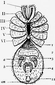
Sub-order a. Mesothelae (see figs. 60 to 62).—Opisthosoma distinctly segmented, furnished with 11 tergal plates, as in the Amblypygi; the ventral surface of the 1st and 2nd somites with large sternal plates, covering the genital aperture and the two pairs of pulmonary sacs, the sternal plates from the 6th to the 11th somites represented by integumental ridges, weakly chitinized in the middle. The two pairs of spinning appendages retain their primitive position in the middle of the lower surface of the opisthosoma far in advance of the anus on the 3rd and 4th somites, each appendage consisting of a stout, many-jointed outer branch and a slender, unsegmented inner branch. Prosoma as in the Mygalomorphae, except that the mesosternal area is long and narrow.
- Family—Liphistiidae (Liphistius, *Arthrolycosa).
Sub-order b. Opisthothelae (see fig. 63).—Opisthosoma without trace of separate terga and sterna, the segmentation merely represented posteriorly by slight integumental folds and the sterna of the 1st and 2nd somites by the opercular plates of the pulmonary sacs. The spinning appendages migrate to the posterior end of the opisthosoma and take up a position close to the anus; the inner branches of the anterior pair either atrophy or are represented homogenetically by a plate, the cribellum, or by an undivided membranous lobe, the colulus.
Tribe 1. Mygalomorphae.—The plane of the articulation of the appendages of the 1st pair to the prosoma (the retrovert) vertical, the basal segment projecting straight forwards at its proximal end, the distal segment or fang closing backwards in a direction subparallel to the long axis of the body. Two pairs of pulmonary sacs.
Families—Theraphosidae (Avicularia, Poecilotheria). Barychelidae (Barychelus, Plagiobothrus). Dipluridae (Diplura, Macrothele). Ctenizidae (Cteniza, Nemesia). Atypidae (Atypus, Calommata). Tribe 2. Arachnomorphae.—The plane of the articulation of the appendages of the 1st pair to the prosoma horizontal, the basal segment projecting vertically downwards, at least at its proximal end, the distal segment or fang closing inwards nearly or quite at right angles to the long axis of the body. The posterior pulmonary sacs (except in Hypochilus) replaced by tracheal tubes; the anterior and posterior pairs replaced by tracheal tubes in the Caponiidae. Principal families—Hypochilidae (Hypochilus). Dysderidae (Dysdera, Segestria). Caponiidae (Caponia, Nops). Filistatidae (Filistata). Uloboridae (Uloborus, Dinopis). Argiapidae (Nephila, Gasteracantha). Pholcidae (Pholcus, Artema). Agelenidae (Tegenaria). Lycosidae (Lycosa). Clubionidae (Clubiona, Olios, Sparassus) Gnaphosidae (Gnaphosa, Hemiclaea). Thomisidae (Thomisus). Attidae (Salticus). Urocteidae (Uroctea). Eresidae (Eresus).
Remarks on the Araneae.—The Spiders are the most numerous


