the exception of the Aplustridae, Lophocercidae and Thecosomata,
the head is devoid of tentacles, and its dorsal surface forms a digging
disk or shield. The edges of the foot form parapodia, often transformed
into fins. Posteriorly the mantle forms a large pallial lobe
under the pallial aperture. Stomach generally provided with
chitinous or calcified masticatory plates. Visceral commissure fairly
long, except in Runcina, Lobiger and Thecosomata. Hermaphrodite
genital aperture, connected with the penis by a ciliated
groove, except in Actaeon, Lobiger and Cavolinia longirostris, in
which the spermiduct is a closed tube. Animals either swim or
burrow.

| |
|
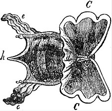
Fig. 49.—Cavolinia tridentata, Forsk. from the Mediterranean, magnified two diameters. (From Owen.) | |
| a, | Mouth. |
| b, | Pair of cephalic tentacles. |
| C, C, | Pteropodial lobes of the foot. |
| d, | Median web connecting these. |
| e, e, | Processes of the mantle-skirt reflected over the surface of the shell. |
| g, | The shell enclosing the visceral hump. |
| h. | The median spine of the shell. |
- Fam. 1.—Actaeonidae. Cephalic shield bifid posteriorly; margins of foot slightly developed; genital duct diaulic; visceral commissure streptoneurous; shell thick, with prominent spire and elongated aperture; a horny operculum. Actaeon, British. Solidula. Tornatellaea, extinct. Adelactaeon. Bullina. Bullinula.
- Fam. 2.—Ringiculidae. Cephalic disk enlarged anteriorly, forming an open tube posteriorly; shell external, thick, with prominent spire; no operculum. Ringicula. Pugnus.
- Fam. 3.—Tornatinidae. Margins of foot not prominent; no radula; shell external, with inconspicuous spire. Tornatina, British. Retusa. Volvula.
- Fam. 4.—Scaphandridae. Cephalic shield short, truncated posteriorly; eyes deeply embedded; three calcareous stomachal plates; shell external, with reduced spire. Scaphander, British. Atys. Smaragdinella. Cylichna, British. Amphisphyra, British.
- Fam. 5.—Bullidae. Margins of foot well developed; eyes superficial; three chitinous stomachal plates; shell external, with reduced spire. Bulla, British. Haminea, British.
- Fam. 6.—Aceratidae. Cephalic shield continuous with neck; twelve to fourteen stomachal plates; a posterior pallial filament passing through a notch in shell. Acera, British. Cylindrobulla. Volutella.
- Fam. 7.—Aplustridae. Foot very broad; cephalic shield with four tentacles; shell external, thin, without prominent spire. Aplustrum. Hydatina. Micromelo.
- Fam. 8.—Philinidae. Cephalic shield broad, thick and simple; shell wholly internal, thin, spire much reduced, aperture very large. Philine, British. Cryptophthalmus. Chelinodura. Phanerophthalmus. Colpodaspis, British. Colobocephalus.
- Fam. 9.—Doridiidae. Cephalic shield ending posteriorly in a median point; shell internal, largely membranous; no radula or stomachal plates. Doridium. Navarchus.
- Fam. 10.—Gastropteridae. Cephalic shield pointed behind; shell internal, chiefly membranous, with calcified nucleus, nautiloid; parapodia forming fins. Gastropteron.
- Fam. 11.—Runcinidae. Cephalic shield continuous with dorsal integument; no shell; ctenidium projecting from mantle cavity. Runcina.
- Fam. 12.—Lophocercidae. Shell external, globular or ovoid; foot elongated, parapodia separate from ventral surface; genital duct diaulic. Lobiger. Lophocercus.

| |
|
Fig. 50.—Shell of Cavolinia tridentata, seen from the side. | |
| f, | Postero-dorsal surface. |
| g, | Antero-ventral surface. |
| h, | Median dorsal spine. |
| i, | Mouth of the shell. |
The next three families form the group formerly known as Thecosomatous Pteropods. They are all pelagic, the foot being entirely transformed into a pair of anterior fins; eyes are absent, and the nerve centres are concentrated on the ventral side of the oesophagus.
- Fam. 13.—Limacinidae. Dextral animals, with shell coiled pseudo-sinistrally; operculum with sinistral spiral; pallial cavity dorsal. Limacina, British. Peraclis, ctenidium present.
- Fam. 14.—Cymbuliidae. Adult without shell; a sub-epithelial pseudoconch formed by connective tissue; pallial cavity ventral. Cymbulia. Cymbuliopsis. Gleba. Desmopterus.
- Fam. 15.—Cavoliniidae. Shell not coiled, symmetrical; pallial cavity ventral. Cavolinia. Clio. Cuvierina.
Tribe 2.—Aplysiomorpha. Shell more or less internal, much reduced or absent. Head bears two pairs of tentacles. Parapodia separate from ventral surface, and generally transformed into


