glistening band, known as the linea splendens; in front of this lies the single anterior spinal artery.
The postero-median fissure (fig. 3, P.) is much deeper and narrower, and has no reflection of the pia mater into it. Where the posterior nerve roots emerge (fig. 3, P.R.) is a depression which is called the postero- lateral fissure, while between this and the postero-median a slight groove is seen in the cervical region, the para median fissure (fig. 3, P.M; see also fig. 2). On looking at fig. 3 it will be seen CVi that the anterior nerve roots (A.R.) do not emerge from a definite fissure.
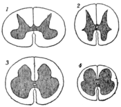
The spinal cord, like the brain, consists of grey and white matter, but, as there is here no representative of the cortical grey matter of the brain, the white matter entirely surrounds the grey. In section the grey matter has the form of an H, the cross bar forming the grey commissure. In the middle of this the central canal can just be made DVn out by the naked eye (see fig. 4). The anterior limbs of the H form the anterior cornua, while the posterior, which in the greater part of the cord are longer and thinner, are the posterior cornua. At the tips of these is a lighter-coloured cap (fig. 3, S.G.) which is known as the substantia gelatinosa Rolandi. On each side of the H is a slighter projection, the lateral cornu, which is best marked in the thoracic region (see fig. 4).
On referring to fig. 4 it will be seen that the grey matter has different and characteristic appearances in different regions of the cord, and it will be noticed that in the cervical and lumbar enlargements, where the nerve to the limbs comes off, the anterior horns are broadened.

(From D. J. Cunningham, in Cunningham's Text-Book of Anatomy.)
Fig. 2.—Diagram of the Spinal Cord as seen from behind.
CVI shows the level of the 1st cervical vertebra; CVv of the 5th cervical vertebra; Dvii of the 2nd dorsal vertebra; DVx of the 10th dorsal vertebra; DVxii of the 12th dorsal vertebra; LVii of the 2nd lumbar vertebra.

Fig. 3.—Diagram to show the Tracts in the Spinal Cord.
A. Antero-median fissure.
P. Postero-median fissure.
A.R. Anterior nerve roots.
P.R. Posterior nerve roots.
P.M. Paramedian fissure.
S.G. Substantia gelatinosa.
G.T. Tract of Goll.
B.T. Tract of Burdach.
C.T. Comma tract.
O.A. Oval area.
L.T. Lissauer's tract.
D.C.T. Direct cerebellar tract.
T.G. Gowers' tract.
C.P.T. Crossed pyramidal tract.
L.B.B. Lateral basis bundle.
A.B.B. Anterior basis bundle.
D.P.T. Direct pyramidal tract.
Histologically the grey matter is made up of neuroglia, medullated and non-medullated nerve fibres, and nerve cells (for details see Nervous System). The nerve cells are arranged in three main columns, ventral, intermedio-lateral and posterior vesicular. The ventral cell column has the longest cells, and these are again subdivided into antero-mesial, antero lateral, postero-lateral and central groups. The intermedio lateral cell column is found in the lateral horn of the thoracic region.
The posterior vesicular or Clarke's column is also largely confined to the thoracic region, and lies in the mesial part of the posterior cornu. It is the place to which the sensory fibres of the sympathetic system (visceral afferents) run. The white matter, as has been shown, surrounds the grey. and passes across the middle line to form the white cornmissure, which lies in front of the grey. It is composed of neuroglia and medullated nerve fibres, which are arranged in definite tracts, although in a section of a healthy cord these tracts cannot be distinguished even with the microscope. They have been and are still being gradually mapped out by pathologists, physiologists and embryologists.
On tracing a sensory nerve into the cord (fig. 3, P.R.) through the posterior nerve root it will be seen to lie quite close to the mesial side of the posterior horn of grey matter, where most of it runs upward. The next root higher up takes the same position and pushes the former one toward the middle line, so that the lower nerve fibres occupy an area close to the postero-median fissure known as the tract of Goll (fig. 3, G.T.), while the higher lie more externally in the tract of Burdach (B.T.). The greater part of each nerve sooner or later enters the grey matter and comes into close relation with the cells of Clarke's column, but some fibres run right up to the nucleus gracilis and cuneatus in the medulla (see Brain), while a few turn down and form a descending tract, which, in the upper part of the cord, is situated in the inner part of the tract of Burdach and is known as the comma tract (fig. 3, C.T.), but lower down gradually shifts quite close to the postero-median fissure and forms the oval area of Flechsig (fig. 3, O.A.). It will be obvious that both these tracts could not be seen in the same section, and that fig. 3 is only a diagrammatic outline of their position.
A few fibres of each sensory nerve ascend in a small area known as Lissauer's tract (fig. 3, L.T.) on the outer side of the posterior nerve roots, and eventually enter the substantia gelatinosa.
To the outer side of Lissauer's tract and lying close to the lateral surface of the cord is the direct cerebellar tract (fig. 3, D.C.T.), the fibres of which ascend from the cells of Clarke's column to the cerebellum. As Clarke's column is only well developed in the thoracic region this tract obviously cannot go much lower.
In front of the last and also close to the lateral surface of the cord is another ascending tract, the tract of Gowers (fig. 3, T.G.), or, as it is sometimes called, the lateral sensory fasciculus. It probably begins in the cells of the posterior horn, and runs up to join the fillet and also to reach the cerebellum through the superior cerebellar peduncle. The crossed pyramidal tract (fig. 3, C.P.T.) lies internal to the direct cerebellar tract, between it and the posterior cornu. It is the great motor tract by which the fibres coming from the Rolandic area of the cerebral cortex are brought into touch with the motor cells in the anterior cornu of the opposite side. This tract extends right down to the fourth sacral nerve.
In front of the crossed pyramidal tract is the lateral basis bundle (fig. 3, L.B.B), which probably consists of association fibres linking up different segments of the cord.
The anterior basis bundle (fig. 3, A.B.B.) lies in front and on the mesial side of the anterior cornu, and through it pass the anterior nerve roots. Like the lateral bundle it consists chiefly of association fibres, but it is continued up into the medulla as the posterior longitudinal bundle to the optic nuclei.
The direct pyramidal tract (fig. 3, D.P.T.) is a small bundle of the motor fibres from the Rolandic area, which, instead of crossing to the other side at the decussation of the pyramids in the medulla, runs down by the side of the antero-median fissure. Its fibres, however, keep on gradually crossing to the opposite side through the anterior white commissure of the cord, and by the time the midthoracic region is reached it has usually disappeared.
The roots of the spinal nerves in the upper part of the canal rise from the cord nearly opposite the points at which they emerge between the vertebrae, but the farther one passes down the higher the origin of each root becomes above its point of emergence. Consequently the lumbar and sacral nerves run a long way down from the lumbar enlargement to their spinal foramina and are enclosed in the dural and arachnoid sheaths to form a mass like a horse's tail, which is therefore known as the cauda equina. The relation between the origin of each nerve and the spinous processes of the vertebrae has been worked out by R. W. Reid (Journ. Anat. and Phys., xxiii. 341).

Fig. 4.—Sections of Spinal Cord, twice scale of nature.
1. Cervical enlargement.
2. Thoracic region.
3. Lumbar enlargement.
4. Sacral region.
Embryology. - The early development of the neural tube from the ectoderm is outlined in the article on the Brain. When the neural groove becomes a tube it is oval in section with a very large laterally
