only within a few months that the true structure of these organs has been made known. Mr. J. D. Hyatt, of this city, has been studying the subject for the past eight years, and his recent discoveries have shown that the ordinary descriptions are incorrect and founded upon mere inferences, drawn from the appearance of the organ as usually dissected and mounted. There are no less than eight discoveries, for which we are indebted to the labors of this gentleman, and it is our intention to present some of these as briefly as possible.
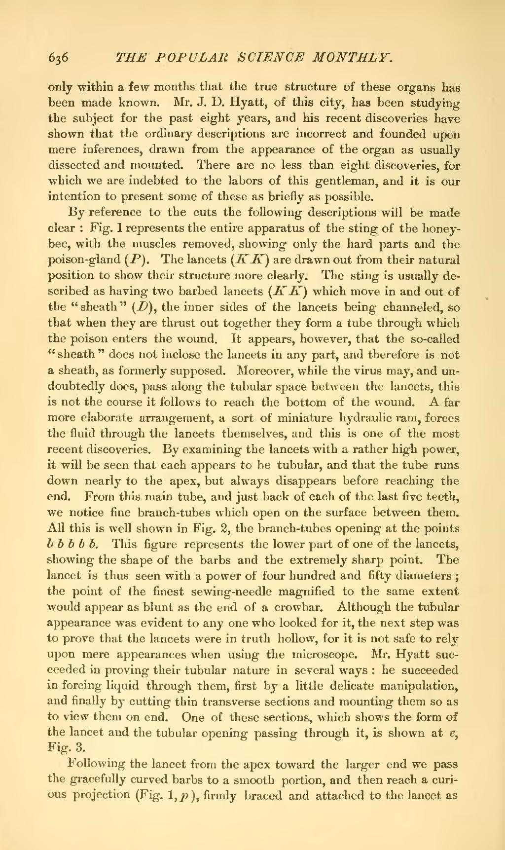
By reference to the cuts the following descriptions will be made clear: Fig. 1 represents the entire apparatus of the sting of the honeybee, with the muscles removed, showing only the hard parts and the poison-gland (P). The lancets (K K) are drawn out from their natural position to show their structure more clearly. The sting is usually described as having two barbed lancets (K K) which move in and out of the "sheath" (D), the inner sides of the lancets being channeled, so that when they are thrust out together they form a tube through which the poison enters the wound. It appears, however, that the so-called "sheath" does not inclose the lancets in any part, and therefore is not a sheath, as formerly supposed. Moreover, while the virus may, and undoubtedly does, pass along the tubular space between the lancets, this is not the course it follows to reach the bottom of the wound. A far more elaborate arrangement, a sort of miniature hydraulic ram, forces the fluid through the lancets themselves, and this is one of the most recent discoveries. By examining the lancets with a rather high power, it will be seen that each appears to be tubular, and that the tube runs down nearly to the apex, but always disappears before reaching the end. From this main tube, and just back of each of the last five teeth, we notice fine branch-tubes which open on the surface between them. All this is well shown in Fig. 2, the branch-tubes opening at the points b b b b b. This figure represents the lower part of one of the lancets, showing the shape of the barbs and the extremely sharp point. The lancet is thus seen with a power of four hundred and fifty diameters; the point of the finest sewing-needle magnified to the same extent would appear as blunt as the end of a crowbar. Although the tubular appearance was evident to any one who looked for it, the next step was to prove that the lancets were in truth hollow, for it is not safe to rely upon mere appearances when using the microscope. Mr. Hyatt succeeded in proving their tubular nature in several ways: he succeeded in forcing liquid through them, first by a little delicate manipulation, and finally by cutting thin transverse sections and mounting them so as to view them on end. One of these sections, which shows the form of the lancet and the tubular opening passing through it, is shown at e, Fig. 3.
Following the lancet from the apex toward the larger end we pass the gracefully curved barbs to a smooth portion, and then reach a curious projection (Fig. l,p), firmly braced and attached to the lancet as