File:EB1911 - Muscular System - Fig 12.png

From Wikisource
Jump to navigation Jump to search

Original file(1,169 × 1,349 pixels, file size: 436 KB, MIME type: image/png)

Summary

Description
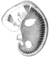
English: Fig. 12.—Scheme to Illustrate the Disposition of the Myotomes in the Embryo in Relation to the Head, Trunk and Limbs.

A, B, C, First three cephalic myotomes.

N, 1, 2, 3, 4, Last persisting cephalic myotomes.

C, T, L, S, Co., The myotomes of the cervical, thoracic, lumbar, sacral and caudal regions.

I., II., III., IV., V., VI., VII., VIII., IX., X., XI., XII., Refer to the cranial nerves and the structures with which they may be embryologically associated.
Date
Source https://en.wikisource.org/wiki/Page:EB1911_-_Volume_19.djvu/73
Author Encyclopædia Britannica

Licensing

Public domain This image comes from the 13th edition of the Encyclopædia Britannica or earlier. The copyrights for that book have expired in the United States because the book was first published in the US with the publication occurring before January 1, 1929. As such, this image is in the public domain in the United States.

Captions

Fig. 12.—Scheme to Illustrate the Disposition of the Myotomes in the Embryo in Relation to the Head, Trunk and Limbs.

Items portrayed in this file

depicts

File history

Click on a date/time to view the file as it appeared at that time.

Date/TimeThumbnailDimensionsUserComment
current11:13, 5 September 2023Thumbnail for version as of 11:13, 5 September 20231,169 × 1,349 (436 KB)Sp1nd01Uploaded a work by Encyclopædia Britannica from https://en.wikisource.org/wiki/Page:EB1911_-_Volume_19.djvu/73 with UploadWizard

Metadata