File:EB1911 - Olfactory System - Fig. 3.png

From Wikisource
Jump to navigation Jump to search

Original file(1,553 × 1,366 pixels, file size: 424 KB, MIME type: image/png)

Summary

Description
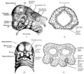
English: Fig. 3.

I. Side view of the head of human embryo about 27 days old, showing the olfactory pit and the visceral arches and clefts (from His).

II. Transverse section through the head of an embryo, showing the relation of the olfactory pits to the fore brain and to the roof of the stomatodaeal space.

III. Head of human embryo about 29 days old, showing the division of the lower part of the mesial frontal process into the two globular processes, the intervention of the olfactory pits between the mesial and lateral nasal processes, and the approximation of the maxillary and lateral nasal processes, which, however are separated by the oculo-nasal sulcus (from His).

IV. Transverse section of head of embryo, showing the deepening of the olfactory pits and their relation to the hemisphere vesicles of the fore-brain.
Date
Source https://en.wikisource.org/wiki/Page%3AEB1911_-_Volume_20.djvu/103
Author Encyclopædia Britannica

Licensing

Public domain This image comes from the 13th edition of the Encyclopædia Britannica or earlier. The copyrights for that book have expired in the United States because the book was first published in the US with the publication occurring before January 1, 1929. As such, this image is in the public domain in the United States.

File history

Click on a date/time to view the file as it appeared at that time.

Date/TimeThumbnailDimensionsUserComment
current13:01, 29 September 2023Thumbnail for version as of 13:01, 29 September 20231,553 × 1,366 (424 KB)Sp1nd01Uploaded a work by Encyclopædia Britannica from https://en.wikisource.org/wiki/Page%3AEB1911_-_Volume_20.djvu/103 with UploadWizard

Metadata