end is played upon by other neurons—spinal, cerebral and
cerebellar.
It is with the neural element of muscle tonus that tendon phenomena are intimately associated. The earliest-studied of these, the “knee-jerk,” may serve as example of the class. It is a brief extension of the limb at the knee-joint, due to a simple contraction of the extensor muscle, elicited by a tap or other short mechanical stimulus applied to the muscle fibres through the tendon of the muscle. The jerk is obtainable only from muscle fibres possessed of neural tonus. If the sensory nerves of the extensor muscle be severed, the “jerk” is lost. The brevity of the interval between the tap on the knee and the beginning of the resultant contraction of the muscle seems such as to exclude the possibility of reflex development. A little experience in observations on the knee-jerk imparts a notion of the average strength of the “jerk.” Wide departures from the normal standard are met with and are symptomatic of certain nervous conditions. Stretching of the muscles antagonistic to the extensors—namely, of the flexor muscles—reduces the jerk by inhibiting the extensor spinal nerve cells through the nervous impulses generated by the tense flexor muscles. Hence a favourable posture of the limb for eliciting the jerk is one ensuring relaxation of the hamstring muscles, as when the leg has been crossed upon the other. In sleep the jerk is diminished, in deep sleep quite abolished. Extreme bodily fatigue diminishes it. Conversely, a cold bath increases it. The turning of attention towards the knee interferes with the jerk; hence the device of directing the person to perform vigorously some movement, which does not involve the muscles of the lower limb, at the moment when the light blow is dealt upon the tendon. A slight degree of contraction of muscle seems the substratum of all attention. The direction of attention to the performance of some movement by the arm ensures that looseness and freedom from tension in the thigh muscles which is essential for the provocation of the jerk. The motor cells of the extensor muscles, when preoccupied by cerebral influence, appear refractory. T. Ziehen has noted exaltation of the jerk to follow extirpation of a cortical centre.
Although the cell body or perikaryon of the neuron, with its contained nucleus, is essential for the maintenance of the life of the cell branches, it has become recognized that the actual process and function of “conduction” in many neurons can, and does, go on without the cell body being directly concerned in the conduction. Conduction in Neurons. S. Exner first showed, many years ago, that the nerve impulse travels through the spinal ganglion at the same speed as along the other parts of the nerve trunk—that is, that it suffers no delay in transit through the perikarya of the afferent root-neurons. Bethe has succeeded in isolating their perikarya from certain of the afferent neurons of the antennule of Carcinus. The conduction through the amputated cell branches continues unimpaired for many hours. This indicates that the conjunction between the conducting substance of the dendrons and that of the axon can be effected without the intermediation of the cell body. But the proper nutrition of the conducting substance is indissolubly dependent on the cell branches being in continuity with the cell body and nucleus it contains. Evidence illustrating this nexus is found in the visible changes produced in the perikaryon by prolonged activity induced and maintained in the conducting branches of the cell. As a result the fatigued cells appear shrunken, and their reaction to staining reagents alters, thus showing chemical alteration. Most marked is the decrease in the volume of the nucleus, amounting even to 44% of the initial volume. In the myelinated cell branches of the neuron, that is, in the ordinary nerve fibres, no visible change has ever been demonstrated as the result of any normal activity, however great—a striking contrast to the observations obtained on the perikarya. The chemical changes that accompany activity in the nerve fibre must be very small, for the production of CO2 is barely measurable, and no production of heat is observable as the result of the most forced tetanic activity.
The nerve cells of the higher vertebrata, unlike their blood cells, their connective tissue cells, and even their muscle cells, early, and indeed in embryonic life, lose power of multiplication. The number of them formed is definitely closed at an early period of the individual life. Although, unlike so many other cells, thus early sterile for Growth in Nervous System. reproduction of their kind, they retain for longer than most cells a high power of individual growth. They continue to grow, and to thrust out new branches and to lengthen existing branches, for many years far into adult life. They similarly possess power to repair and to regenerate their cell branches where these are injured or destroyed by trauma or disease. This is the, explanation of the repair of nerve trunks that have been severed, with consequent degeneration of the peripheral nerve fibres. As a rule, a longer time is required to restore the motor than the sensory functions of a nerve trunk.
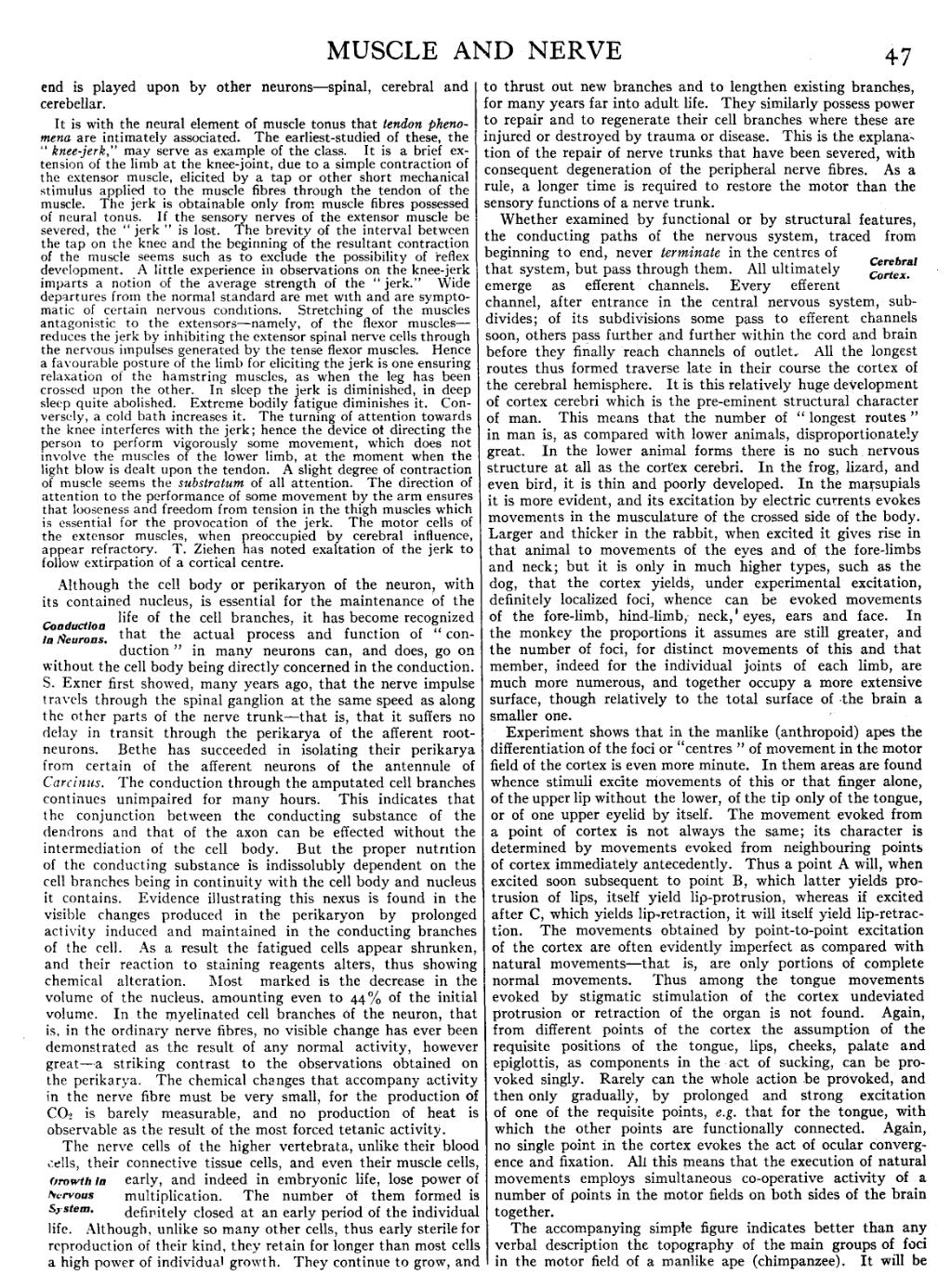
Whether examined by functional or by structural features, the conducting paths of the nervous system, traced from beginning to end, never terminate in the centres of that system, but pass through them. All ultimately emerge as efferent channels. Every efferent channel, after entrance in the central nervous system, subdivides; Cerebral Cortex. of its subdivisions some pass to efferent channels soon, others pass further and further within the cord and brain before they finally 'reach channels of outlet. All the longest routes thus formed traverse late in their course the cortex of the cerebral hemisphere. It is this relatively huge development of cortex cerebri which is the pre-eminent structural character of man. This means that the number of “longest routes” in man is, as compared with lower animals, disproportionately great. In the lower animal forms there is no such nervous structure at all as the cortex cerebri. In the frog, lizard, and even bird, it is thin and poorly developed. In the marsupials it is more evident, and its excitation by electric currents evokes movements in the musculature of the crossed side of the body. Larger and thicker in the rabbit, when excited it gives rise in that animal to movements of the eyes and of the fore-limbs and neck; but it is only in much higher types, such as the dog, that the cortex yields, under experimental excitation, definitely localized foci, whence can be evoked movements of the fore-limb, hind-limb, neck, eyes, ears and face. In the monkey the proportions it assumes are still greater, and the number of foci, for distinct movements of this and that member, indeed for the individual joints of each limb, are much more numerous, and together occupy a more extensive surface, though relatively to the total surface of the brain a smaller one.
Experiment shows that in the manlike (anthropoid) apes the differentiation of the foci or “centres” of movement in the motor field of the cortex is even more minute. In them areas are found whence stimuli excite movements of this or that finger alone, of the upper lip without the lower, of the tip only of the tongue, or of one upper eyelid by itself. The movement evoked from a point of cortex is not always the same; its character is determined by movements evoked from neighbouring points of cortex immediately antecedently. Thus a point A will, when excited soon subsequent to point B, which latter yields protrusion of lips, itself yield lip-protrusion, whereas if excited after C, which yields lip-retraction, it will itself yield lip-retraction. The movements obtained by point-to-point excitation of the cortex are often evidently imperfect as compared with natural movements—that is, are only portions of complete normal movements. Thus among the tongue movements evoked by stigmatic stimulation of the cortex undeviated protrusion or retraction of the organ is not found. Again, from different points of the cortex the assumption of the requisite positions of the tongue, lips, cheeks, palate and epiglottis, as components in the act of sucking, can be provoked singly. Rarely can the whole action be provoked, and then only gradually, by prolonged and strong excitation of one of the requisite points, e.g. that for the tongue, with which the other points are functionally connected. Again, no single point in the cortex evokes the act of ocular convergence and fixation. All this means that the execution of natural movements employs simultaneous co-operative activity of a number of points in the motor fields on both sides of the brain together.
The accompanying simple figure indicates better than any verbal description the topography of the main groups of foci in the motor field of a manlike ape (chimpanzee). It will be