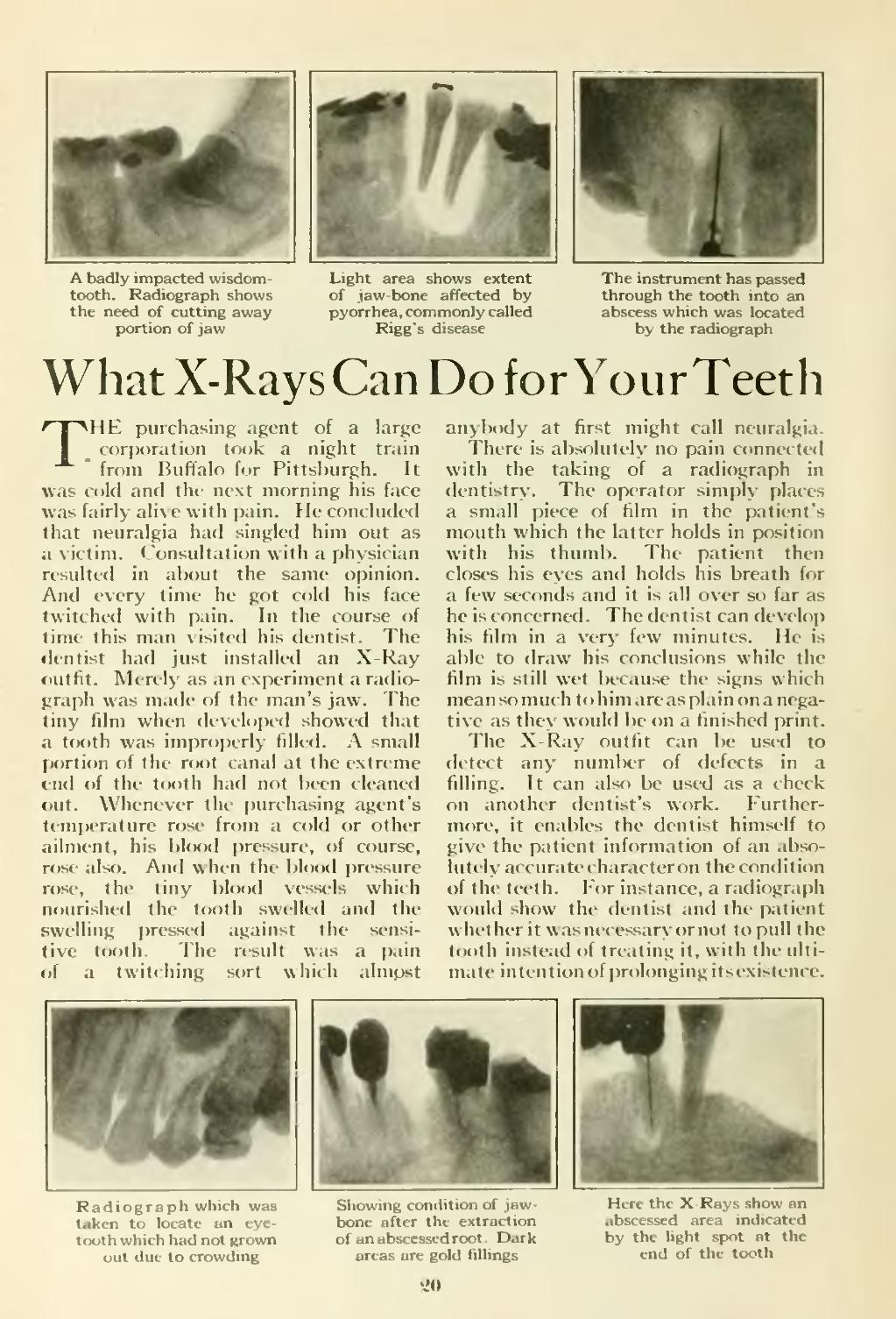
��A badly impacted wisdom- tooth. Radiograph shows the need of cutting away portion of jaw
��Light area shows extent
of jaw-bone affected by
pyorrhea, commonly called
Rigg's disease
��The instrument has passed
through the tooth into an
abscess which was located
by the radiograph
��What X-Rays Can Do for Your Teeth
��THE purchasing agent of a large corporation took a night train ' from BufTalo for Pittsburgh. It was cold and the next morning his face was fairly alive with pain. He concluded that neuralgia had singled him out as a victim. Consultation with a physician resulted in about the same opinion. And every time he got cold his face twitched with pain. In the course of time this man visited his dentist. The dentist had just installed an X-Ray outfit. Merely as an experiment a radio- graph was made of the man's jaw. The tiny film when developed showed that a tooth was improperly filled. A small portion of the root canal at the extreme end of the tooth had not been cleaned out. Whenever the purchasing agent's temperature rose from a cold or other ailment, his blood pressure, of course, rose also. And when the blood pressure rose, the tiny blood vessels which nourished the tooth swelled and the swelling pressed against the sensi- tive tooth. The result was a pain of a twitching sort which almcist
��anybody at first might call neuralgia.
There is absolutely no pain connected with the taking of a radiograph in dentistry. The operator simply places a small piece of film in the patient's mouth which the latter holds in position with his thumb. The patient then closes his eyes and holds his breath for a few seconds and it is all over so far as he is concerned. The dentist can develop his film in a very few minutes. He is able to draw his conclusions while the film is still wet because the signs which mean so much to him are as plain on a nega- tive as they would be on a finished print.
The X-Ray outfit can be used to detect any number of defects in a filling. It can also be used as a check on another dentist's work. Further- more, it enables the dentist himself to give the patient information of an abso- lutely accurate character on the condition of the teeth. For instance, a radiograph would show the dentist and the patient whether it wasnccessaryornol to pull the tooth instead of treating it, with the ulti- mate intention of i)r()longing its existence.
���Radiograph which was Showinf cnmlition of jaw- taken to locate an eye- born iii ilu extraction tooth which had not grown ofjm i ■ ..Iroot. Dark out due to crowding in ild fillings
��Here the X Rays show an
abscessed area indicated
by the light spot at the
end of the tooth
��•vdO
�� �