Popular Science Monthly/Volume 6/March 1875/Biology for Young Beginners II
| BIOLOGY FOR YOUNG BEGINNERS.[1] |
By SARAH HACKETT STEVENSON.
II.
IN the chimney-corner by the kitchen-fire stood a quaint stone jar that every winter morning bubbled over with the light, gray foam of buckwheat-cakes. While our "mouths watered," our minds wondered—wondered at the magic by which so many cakes were made out of so little flour. We believed there were fairies in the yeast; but it was only the other day that I succeeded in finding these fairies, and I want to tell you how you may find them too.
To begin with, you must have a spoonful or two of yeast to look at while I talk. You will probably notice first a number of bubbles, like soap-bubbles; but, instead of common air, the yeast-bubbles contain a gas made by the yeast—carbonic-acid gas. Next you will notice the brownish color of the yeast; it grows thicker and muddy, and after an hour or so begins to rise. This rising the chemists call fermentation; biologists call it growing. The spoonful has become a cupful. The yeast is really alive, and it is one of the simplest forms of life. In studying biology, then, or the science of life, we begin at what seems the beginning.
All that I have described you can see with your own eyes; but now I must tell you something about the yeast which you could never find out with your eyes alone. With the aid of the microscope, a great many little solid bodies are seen floating about in it. Sometimes they are found alone, but most frequently in groups (Figs. 1-5),
| Fig. 1.—Round Cells. | Fig. 2.—Lemon-shaped Cells. | Fig. 3.—Outer and Inner Surface of Sac. |
 | |
| Fig. 4.—Group of Cells. | Fig. 5.—Group of Cells. |
and each one is about 13000 of an inch in size. Though they are solid, yet we can see through them, and they are always round (Fig. 1), some of them not quite as round as a ball, more like a lemon (Fig. 2), but none of them are square or flat. The cover of each one is double (Fig. 3), that is, it has an outside and an inside. Under the microscope, these two surfaces look like two round lines, one within the other (Fig. 6). Inside these lines is something which looks like little grains (Fig. 7); and this whole cover, with all that is inside of
 | |||
| Fig. 6. | Fig. 7. | Fig. 8. | Fig. 9.—Yeast-cell, or Torula. a, Jelly, or Protoplasm; |
it, is called a cell. Now you must learn of what these cells are made. First there is the outside part, which is like a bag or sac. This bag is tough and solid, and is full of a jelly-like substance, which is thick and brownish, next the wall of the bag, but thinner and more transparent toward the centre (Fig. 8). This jelly (a) is called protoplasm, and the thin space (b) in the centre is an air-cell or vacuole (Fig. 9). If you color the yeast-cells, you can see the different parts much better. A drop of magenta will pass right through the sac without staining it at all. The cell-jelly, or protoplasm, will be quite red, and the vacuole will not be colored, though it may look pinkish, because you see it through a layer of the protoplasm (Figs. 10, 11). Now, if the cell were all made of the same material, it would probably all be colored by the magenta.
These cells are called torulæ—a single one is a torula. The word means a little knobby swelling. You will soon see how it comes to
 | ||
| Fig. 10.—Jelly stained, and the Sac clear. | Fig. 11.—Broken Cell.Sac clear, Jelly stained. | |
have this name. If you have followed me carefully, better still it you have seen it all for yourselves under the microscope, you know that the torulæ are alive, and that they grow. Every thing that grows must have food. Now, whence does the torula get its food? From the liquid in which it floats. What is this liquid? The greater part is water, but if you sow yeast in pure water it will hardly grow at all; but if you put in ever so little sugar, it will froth and bubble considerably. If besides the sugar you give it the least bit of ammonia, magnesia, lime, and potash,[2] it will thrive splendidly. The torula takes in this food, and churns it up into that "elixir of life" or protein, woody cells or cellulose, and fat. Then, if you watch carefully, you will see a whole lot of little buds coming out around the edges of the wall (Fig. 12); hence the torula is really a little knobby swelling. Some of the buds have other buds at their edges; all these buds are the little baby-torulæ. By-and-by they break away from the old mother-torula, but they always pay visits back and forth, and sometimes build their houses right next the parental roof in clusters (Fig. 13); at other times they build in long rows, like a chain or a string of beads (Fig. 14). Of this you may be sure, every torula has a mother. People have been trying to prove for two hundred years or more that these little specks of life can make themselves. Some time I will tell you how you can prove that this is not true. These little torulæ float about in the air, or sleep in any dry place, never showing that they are alive till they are planted in some nest or nidus. When the cook dries her yeast-cakes, she puts all the little torulæ to sleep, and there they go into winter quarters, or hibernate in their cells, like the bears in their caves.
 | ||
| Fig. 12.—Cell and its Buds. | Fig. 13.—The Baby Torulæ grouped around their mother. | Fig.—14.—Cells linked together in Chains. |
There is still another appearance of yeast. Let your cup of yeast stand long enough, and do not add any more sugar or water to it, you will find that the bubbling or fermentation stops, the torulæ settle to the bottom, and the fluid comes to the top. This fluid has a strong or biting instead of a sweet taste, like the fluid into which you first placed the yeast. The fermentation has changed its nature—the fairy torulæ with their magic wands have turned the sugar into carbonic acid, alcohol, glycerine, and succinic acid. These are called the products of fermentation. The carbonic acid, you know, passed off through the bubbles, the other products are still in the fluid. A little of this fluid will make you merry; if you take much of it, you will become intoxicated. This is due to the alcohol, and the value of yeast depends upon its power to make alcohol. You may know that the fluid is alcohol if, when you touch it with a lighted match, it burns with a blue flame.
Now, I have told you the torula grows—it has life; but how does it grow—as a mineral, a vegetable, or an animal? The mineral grows larger and larger by additions made to its outside. This is called growth by accretion. But the torula or yeast-cell grows by taking in new substance in among the particles of its old substance, and this kind of growth is called by a long name—intussusception. This is one of the reasons why it is not a mineral. Is it an animal? The line that divides the animal from the vegetable kingdom is not very well marked, but there are two reasons why the torula is not an animal. In the first place, its jelly or protoplasm is shut up in a close sac, but the protoplasm-jelly of animal cells forms a wall of itself. In the second place, the torula can make its own food or protein out of the raw material it finds in the liquid, while the animal cells seem to have no such power; they must have their protein already made, and their work is to destroy it. So, if the torula is not a mineral nor an animal, it must be a vegetable. Vegetables are the manufacturers or producers of protein; animals are the destroyers or consumers of it.[3] You have now found that the torula or yeast-cell is a plant, and not an animal. The next question is, What kind of a plant is it? Mostly all plants need the sun, but the yeast-plant grows as well in the dark as in the light. Plants that need the light are always green; they take in carbonic acid, and give off oxygen, but the torula has no green color, and it takes in oxygen and gives off carbonic acid. Those plants which give off carbonic acid, grow in the dark, and are not green, are called fungi. The mushrooms and toadstools are fungi. Now let us see how many things you have learned about yeast: First, that it is alive; second, that it is a plant; third, that it is a fungus.
When first you try to study this lobster, you will perhaps think, as I thought, "How can I straighten out such a queer, crusty, clawy thing as that?" But, though the lobster looks as hard as the Greek alphabet, he is as easy as your own A, B, C, when once you find him out. You know the corolla or crown of the bean looked so hard, but it all came out nicely into five leaves, or petals, as soon as you knew how. Now let us see if we can find and name the different parts of the lobster. (You must have a real lobster before you to look at while I talk. The crawfish or crayfish that lives in brooks and rivers is fashioned after the lobster, only smaller; so one of these can be studied by those of you who live inland.) One thing is very certain—he has a great many different parts, very unlike each other. First, you see (Fig. 15), he is covered with a shell, which, like the mussel's and clam's, is his exo-skeleton. This shell is very hard, like stone, and it is colored purplish black with pale spots here and there. The lobsters which you see in shops are always scarlet. When these poor fellows are caught, they are plunged alive into boiling water, which turns the black coat red. This outside shell or exo-skeleton is made up of a great many different pieces, instead of two, as the mussel's; but those pieces are shaped and joined in such a way as to make three divisions of the body—a head, a thorax, or breastplate, and an abdomen. The head-piece of the shell is pointed in front, forming the beak or frontal spine (Fig. 15). Behind this head-piece is a groove or seam where the head joins the breast or thorax, making the two pieces of shell which cover the head and breast all one. So the first and second divisions of the body thus joined in one are called the thorax or head breastplate. The large piece of shell, with the seam that covers the back and sides of the cephalo-thorax, is called the carapace or shield (Fig. 15). It is the front sharp point of this shield (carapace) that is called the frontal spine or beak. Behind the head and breast (cephalo-thorax) lies the third division of the body—the

Fig. 15
abdomen—which is made up of seven pieces or joints. The first six joints are called somites (Fig. 15) or bodies, and the last joint or tail-piece is called a telson, which means end. So the body of the lobster is made up of six somites and a telson. Each body-piece has a pair of soft-jointed paddles on its under side (Figs. 15, 16), and these are called swimmerets or little swimmers. The lower joints of these paddles have two broad, flat toes. The paddles on the last or sixth somite are different from the others; they are wider and turned backward (Fig. 15) so as to lie at each side of the tail-piece, telson; and these great-fingered paddles, taken with the telson, form what is called the tail-fin. The under or ventral part of each somite, which lies between the paddles, is called the sternum. The rounded upper or dorsal part of the body-piece is the tergum, which means the back. In front of the abdomen, with its somites, is the cephalo-thorax. This cephalo-thorax has a tergum, or back part, a sternum, or under part, a pleuron, or side part (Fig. 15), and so many things

Fig. 16.—One of Lobster's Body-pieces, or Somites.
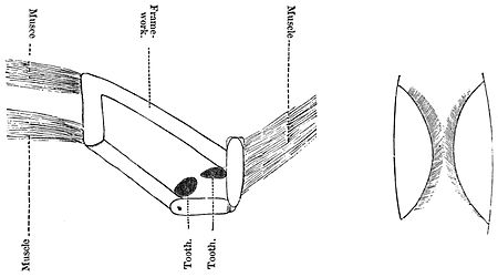
are hanging down from it one can hardly count, much less learn them. Counting from behind forward, you will find between the lobster's body, or abdomen, and the head, eight pair of jointed legs, one pair much longer and larger than the others, with huge pincers at the ends. All these eight pair are called the thoracic appendages, because they are fastened to the thorax, or breastplate. The lobster uses the four back-pairs for walking, and so they are called the ambulatory limbs. The last pair has seven joints, and every joint works in a different direction; so, when these hind-legs start off, it is hard to tell where they intend to go. The next pair of walking-legs are like the hindmost pair, except that the first joint sends out a piece above it, which is kept out of sight in a little room in the side of the lobster (Fig. 22). We shall say more about this room by-and-by. The two front pair of walking-legs send up pieces also into this chamber, but the end of the leg is different from the last two pairs, for they have pincers, or chelæ. Now we have come to the largest pair; the chelæ, or pincers at the ends, are so large and strong that they are called the "great chelæ." They are the lobster's weapons of defense. When he is taken prisoner, that is, seized by one of his claws, he quietly leaves the claw in the hands of his astonished captor, and beats his retreat as fast as possible. He has another odd way of laying down his arms when he is frightened by a great noise, such as thunder, or the tiring of a cannon. It is no uncommon thing to find a number of these broken swords lying about among the rocks, showing where there has been a lobster fright or fight. As soon as one claw goes, another takes its place, but it is some time before the new one gets as long and strong as the old one. You will notice quite a difference between the two large claws, or forceps. In one, the teeth are large and blunt (Fig. 17), and in the other they are very sharp (Fig. 18).
 | |
| Fig. 17.—Large-toothed Claw. | Fig.—18. Small-Toothed Claw. |
The blunt-toothed pincers the lobster uses as an anchor to moor himself, while with the other he attacks and seizes his prey. So much for the great claws, or chelaæ. The next three pair are called maxillipedes, or foot-jaws, because they act both as teeth and feet (Fig. 15). The hindmost foot-jaw has three divisions. One branch passes up into the side-chamber of the lobster; the middle branch is long and jointed: this, and its fellows on the other side, act as a pair of scissors, cutting the food. The third branch is jointed, and is a walking-leg. The middle foot-jaw (maxillipede) is much like the last, while the front one does not send a piece upward into the side-chamber (Fig. 22), and one of its branches is flattened out, so as to look like leaves. The four walking-legs, the great pincers (chelæ) and the three pair of foot-jaws (maxillipedes), making eight pair in all, belong to the lobster's breast (thorax). Now we come to the head, which is provided with six pair of "hangers-on," or appendages. The two back-pair belonging to the head are called maxillæ, because they lie at the side of the mouth, and are like jaws. The hindmost of the jaws—or maxillæ—on each side has a boat-shaped, or oval plate (Fig. 22), which lies at the front entrance of the side-chamber, about which we will hear more presently. The ends of the front pair of little jaws (maxillæ) are leafy, like those of the front pair of foot-jaws (maxillipedes). Now we come to the jaw itself, or mandible, which has strong teeth, bears a small appendage, the palp, and lies at the side of the mouth. From all this you see that the mouth of the lobster is well armed with teeth and scissors to tear and cut its food. Counting from the front, it has first the true jaws (mandibles); then the two pair of little jaws (maxillæ); and these are followed by the three pair of foot-jaws (maxillipedes)
 | |
| Fig. 19.—Long Feeler, or Antenna. | Fig. 20.—Little Feeler, or Antennule and Ear-tube. |
making, altogether, six pair, which are all turned up against the mouth. In front of the jaw are two very long jointed feelers called antennas, but you seldom see them at their full length (Fig. 15); they are easily broken (Fig. 19). Next to the feelers (antennæ) are two

Fig. 21. Eye and Eye stalk.
little feelers, or antennules (Figs. 15, 20); and last of all, in front, comes a pair of joints which support the eyes (Fig. 21), called the optic pair of appendages. Now let us begin with the eyes, and go back to the tail, to see how many pairs of feelers, jaws, hands, feet, and paddles the lobster owns (Fig. 15). He has six pairs attached to the head, eight pair to the breast (thorax), and six pairs to the body (abdomen); in all, twenty pairs, and very few of these appendages are alike.
NAMES OF LOBSTER'S APPENDAGES.
| Head appendages. | I | Pair.... | Eye-stalks. | ||||
| II | ".... | Antennules, or small feelers. | |||||
| III | ".... | Antennae, or great feelers. | |||||
| IV | ".... | Mandibles, or true jaws. | |||||
| V | ".... | Maxillæ, or little jaws. | Firstpair | ||||
| VI | ".... | Second" | |||||
| Thoracic appendages. | VII | ".... | Maxillipedes, or foot-jaws | First" | |||
| VIII | ".... | Second" | |||||
| IX | ".... | Third" | |||||
| X | ".... | Chelæ, or pincers | |||||
| XI | ".... | Ambulatory limbs, or walking legs | XI and XII | ||||
| XII | ".... | with pincers | |||||
| XIII | ".... | XIII and XIV | |||||
| XIV | ".... | without pincers | |||||
| Abdominal appendages | XV | ".... | Swimmerets, or little swimmers. | ||||
| XVI | ".... | ||||||
| XVII | ".... | ||||||
| XVIII | ".... | ||||||
| XIX | ".... | ||||||
| XX | ".... | ||||||
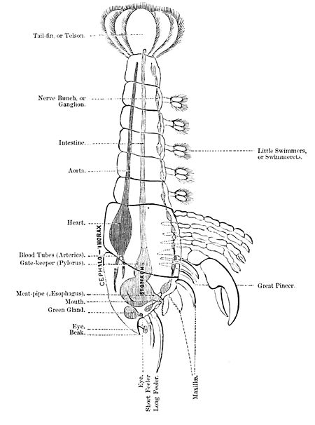
You now have a pretty good idea of the exo-skeleton, or hard outside part of the lobster, and we shall look next at the soft parts inside (Fig. 22). The mouth seems a very good place to begin at, and you will find it between the mandibles, or jaws. In front of it is a lip, shaped like an escutcheon, and is called the labrum, which means lip. At the back of the mouth is another lip, the metastoma, meaning beyond the mouth, and this is looked upon as the lower lip. The mouth, as in the mussel, opens into a gullet, or œsophagus. This meat-pipe opens into a four-cornered box (Fig. 22)—the stomach—which is very curiously made.
Near the centre of the box the walls come almost together, dividing it into two parts: the front part is the larger, and it is called the cardiac end, because in the human body the first part of the stomach points toward the heart, but you see, in the lobster, it points away from the heart. It contains three strong, colored teeth, fastened to a T-shaped frame (Fig. 23), and worked by muscles which are fastened to the inside of the breastplate (carapace). These teeth meet in the middle of the stomach, and form a powerful grinding-machine, which crushes the food like the stones in a mill (Fig. 24). Sometimes, when you find the empty shell of a lobster on the sea-shore, you can see a perfect mould of the old mill—"the mill-wheel gone to decay." How the lobster sets out of his shell, and how he turns the mill out of his stomach, we shall study after a while. The small back part of the stomach is called the pyloric end, and it is made inside like a sieve or strainer. The sides are stuffed out in the centre like cushions, and quite covered with hairs (Fig. 25). Let us see why. Pylorus means gate-keeper. It protects or guards the intestine from all intruders,

Fig. 22.
such as big pieces of meat and hard bodies. None but the finest particles can pass through the strainer, and hence this pylorus is a very good gate-keeper. The intestine does not go wandering about in the body like the mussel's, but passes straight back (Fig. 22), and ends at the anus, at the under part of the tail-piece (telson). On each side of the cephalo-thorax lies a long, soft, yellowish-green mass. This is the liver, and it opens into the small, pyloric end of the stomach by several ducts or pipes on each side. Away up in the front part of the cephalo-thorax, at the base of the feelers (antennæ) on either side

Fig. 23.—The Lobster's Mill.
you may see a soft green mass called the "green gland" (Fig. 22). This is supposed to be the kidney. Next we will take a look into the side-chambers (Fig. 22) of the cephalo-thorax, and see what the three pair of walking-legs, the great pincers (chelæ), and the two pairs of jaw-feet, are doing in there. In each chamber we find eighteen little,
 | |
| Fig. 24.—The Mill and its Muscles. | Fig. 25.—The Hairy Cushions at the small end of the stomach, through which the Food is strained. |
tapering, feathery-like bodies. Each has a central stem, surrounded by fine, feathery filaments. They look very much like so many little bottle-brushes (Fig. 22). These are the gill-plumes, and this room is called the gill, or branchial chamber. The gills are placed in two sets, six in one and twelve in the other. The first row is fastened to the six feet or appendages of the breast (thorax) which we found pushing themselves up into the chamber (Fig. 22). The other twelve are fastened to the pleuron or side-pieces of the cephalo-thorax. These gills are not covered with stiff hairs (cilia) as the mussel's, so there must be some other plan of moving the water. There is a very curious piece of machinery at the front entrance. You remember the oval or boat-shaped plate in front of the chamber, formed by the hind-most little jaw (maxilla). This plate is called the scapho-gnathite, which means the little skiff-like jaw. It is made on the plan of the Archimedean screw, and it works as the screw of a propeller, and is set in motion by the jaws. The water enters the back part of the gill-chamber by a slit, and it is scooped out by the screw through the opening in front, bubbling and frothing as it goes. Thus the mechanism of the screw was all worked out in our little lobster long years before it was discovered by the great Archimedes. The tiny network of the blood-vessels is spread over the framework of the gill-plumes, just as you found it on the lattice-work of the mussel's gill-pockets. As the screw propels the water through the branchial or gill chamber the blood takes out the oxygen from the air in the water, and gives back carbonic acid. You remember how the strong hairs (cilia) of the pockets sweep the water along over the mussel's gills, and how the little blood-vessels take up their oxygen and give up their carbonic acid. The gills that are fastened to the legs move when the legs move, and the faster they go the more water they use. So much for the lobster's breathing or respiration. We will leave his circulation, his muscles and nerves, for another chapter.
 | ||
| Fig. 26. | Fig. 27.—The Lobster's Eye, showing now the Nerve is joined to the Windows in front by Rods and Cones. | |
The eyes, as you have seen, are away in front (Figs. 15, 22) at the ends of the first pair of appendages—the eye-stalks. The eye is kidney-shaped; instead of having one window or pupil as your eye has, through which the light enters, the whole front is divided into squares like old-fashioned window-panes (Fig. 26). Each square is really a separate eye, and this is what is called a compound eye. The lobster's eyesight must be very good, for, besides having all those eyes, the stalks are jointed so that he can turn them in different directions. The nerve which goes to the eye is called the optic nerve, and it is connected with each square by pretty rods and cones, which look like those in your own eye (Fig. 27). The rods and cones are covered with coloring-matter or pigment, which turns red when it is boiled. The optic nerve is a nerve of sensation, because it gives the lobster the sense of sight.
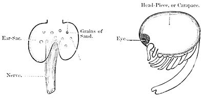
Now, where are the lobster's ears? Not in the foot, as the mussel's, but in their proper place—the head. If you look at the base of the little feelers, on each side you will find a little three-cornered slit, covered with hairs (Fig. 28). This slit leads into a small sac filled
 | ||
| Fig. 28.—Little Feeler, or Antennule, showing Ear-Tube at its Base. | Fig. 29.—The Lobster's Ear. | |
with water. One side of this sac is pushed inward to form a sort of fold or pocket, in which a nerve which comes from the brain or head—ganglia—spreads itself out (Fig. 29). The side of the pocket toward the water is covered with fine hairs, and these hairs touch against little bits of sand which get into the water through the outside slit (Fig. 30). These particles of sand are like the tiny stones or otoliths you found in the mussel's ear-sac, and they, likewise, help to increase the sound. The lobster's ear is made on much the same plan as your own; the sac is really a fold of the lobster's skin, which is pushed in as you might push in the crown of your soft hat. Now, I dare say you are wishing to hear about the lobster's bairns or little ones. The lobster's eggs are covered by a soft, sticky glue, which fastens them to the long hairs which cover the paddles under the abdomen (Fig. 22). The good mother-lobster doubles up her body so that the eggs are all folded inward safe from harm. Hundreds of eggs are carried in this way, and when the lobster is boiled they turn red and form what is called the coral. The baby-lobsters differ greatly from their parents. Their eyes are very large, and set in the head instead of on eye-stalks. They have a great rounded head-shield (carapace) and a small body (Fig. 31). The limbs are not at all like the lobster's; altogether, he looks as if his eyes and head were running away with him. As soon as he is hatched he begins to swim about and feed himself, and never
 | |
| Fig. 30. | Fig. 31.—Young Lobster. |
goes back to the old home. Of course, as he grows, his shell gets too small, but, instead of putting on an addition as the mussel does, he leaves the old house altogether and builds a new one. In three days after the lobster moves out of the old house he has been found all settled in a bran-new one one-third larger. Two round balls are often found in the lobster's stomach, and people call them "crab's-eyes." These balls are made of lime, which it is said the lobster has been storing up for his new shell. Thus the lobster moves "out of the old house into the new" every year till he gets his growth. Then he lives contentedly under the same roof till he dies, or till some one throws him into a lobster-pot.
- ↑ From "Boys and Girls in Biology," in the press of D. Appleton & Co.
- ↑
1pasteur's fluid. Parts. Parts. Potassium phosphate . . . . 20 Cane-sugar . . . . . . . . . . . . 1,500 Calcium phosphate . . . . . . 2 Water . . . . . . . . . . . . . . . . 8,376 Magnesium sulphate . . . . . 2 10,000 Ammonium tartrate . . . . . . 100 - ↑ Such plants as the Venus fly-trap seem to be an exception to this demarcation between the two kingdoms. These plants really digest protein matter, being supplied with what may be termed prehensile organs for capturing their prey. Dr. Hooker suggests that these plants are not exceptional and singular; that they simply continue through life the process begun by the germ when it nourishes itself upon the ready-made food stored up in the seed.


